AI-Driven Segmentation on Weight Bearing CT Introduction Accurate segmentation is critical for orthopedic workflows, including…
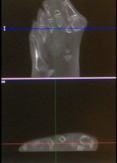
pedCAT: Visualizing the Sesamoids in a Hallux Valgus Deformity

Dr. Robert Weinstein, DPM, recently lectured at The International Foot & Ankle Foundation for Education and Research meeting in Maui, Hawaii, and presented an illuminating finding from his clinical practice: the pedCAT can be a useful diagnostic tool for a very common foot condition.
When it comes to Hallux Valgus, or bunions, a 3D scan could make a major difference in the surgical plan.
Valgus, by definition is a three-dimensional problem, since there is a rotational component, Dr. Weinstein explained.
And “sesamoids, in my mind, drive the whole deformity,” Dr. Weinstein said. “You have to be able to look at them very carefully.”
However, the standard positioning for the “sesamoid view” on plain X-Ray places the patient in an abnormal position, and therefore is not clinically relevant, he added.
“Sesamoid condition is just as important as sesamoid position,” Dr. Weinstein said. “If I see sesamoids that are diseased, it doesn’t matter how good I am at reconstructing those MPJs, that I can get that metatarsal back to zero or two degrees, it’s going to be a failure. So it’s very important to look at the crista and the condition of each of those sesamoids.”
Dr. Weinstein practices at the Ankle & Foot Centers of Georgia.
Dr. Matthew Welck, MD, FRCS, of the Royal National Orthopedic Hospital in London, presented on a similar topic at the AOFAS/IFFAS Annual Meeting in Chicago in a lecture titled, “The Metatarsosesamoid ‘The Empty Crista Sign’ – Could This be a Predictor of Deformity Recurrance after Hallux Valgus Surgery?”
Related Posts