Fragility fractures are often the first visible sign of underlying osteoporosis but too often, they…
Case Study: pedCAT vs. Radiographs
Surgeons often make assumptions based upon plain radiographs. But plain radiographs often hide or distort significant radiographic findings due to bony superimposition.
Take, for example, the case below:
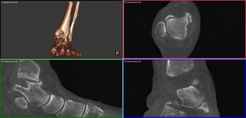
A patient sought a second opinion for the cause of her medial foot and ankle pain. The treating physician used plain radiographs to diagnose her with posterior tibial tendon dysfunction/partial tear and degenerative joint disease of the 1st, 2nd, and 3rd tarsometatarsal joints. The treating physician also noted a “chip” of bone on the inside of the ankle.
The treating physician planned on performing a flat foot reconstruction, a posterior tibial tendon repair, and a tarsometatarsal joint fusion.
The physician performing the second opinion noted the patient’s discomfort over the medial ankle gutter was much more significant than over the posterior tibial tendon and the spring ligament. The patient had minimal discomfort through the tarsometatarsal joints. The physician performing the second opinion ordered a weight bearing pedCAT study to assess the midfoot DJD and to better evaluate the midtarsal joint and ankle joint. The pedCAT study clearly documented a degenerative process in the medial ankle gutter with a bony impingement. On secondary exam, the majority of the symptoms arose from the medial ankle gutter.
If the flat foot reconstruction was performed as planned, the talus would have been dorsiflexed and the tibio-talar impingement would have been worsened. The pedCAT images helped prevent an un-necessary surgery, while directing the physician to the appropriate pathology.

To learn how more about how the pedCAT could benefit your patients, contact CurveBeam today.
Related Posts