Orthopedic decision-making depends on accurate representation of anatomy—particularly when joint alignment and bone relationships change…

Weight Bearing CT Scans for the Evaluation of Implant Arthroplasty Candidates
Weight bearing CT scans can be critical to a proper diagnosis, even for routine procedures.
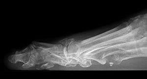
In the following case, for example, a patient’s X-Rays indicated that he would be a good candidate for a metatarsal head hemi-implant arthroplasty. However, when the patient sought a second opinion, a weight bearing CT (pedCAT) scan revealed the true condition of the metatarsal head, and the surgical plan was considerably altered as a result.
A 60 year-old male presented complaining of a many year history of 2nd metatarsophalangeal joint pain, especially joint stiffness and pain. His pain increased with attempted 2nd MTPJ dorsiflexion. In gait, he felt pain when rolling onto the ball of the foot. His first surgical opinion recommended a metatarsal head hemi-implant arthroplasty .
Due to the excessive bony superimposition on the patient’s lateral X-Ray, it is difficult to accurately assess the shape of the 2nd metatarsal head. A bone fragment can be visualized over the dorsum of the first or second metatarsal heads. The AP weight bearing images demonstrate 2nd metatarsophalangeal joint space narrowing.
The patient sought a second opinion from a podiatric surgeon who offers in-office weight bearing CT services. The podiatrist performed a pedCAT scan and found the 2nd metatarsal head had sustained an old fracture. The pedCAT scan revealed that the dorsal 50% of the 2nd metatarsal head had been avulsed dorsally and a portion of the metatarsal head presented as a dorsal loose body. The 2nd metatarsal head didn’t have the bone stock or bone volume to support a hemi-implant. The second opinion recommended recontouring the metatarsal head and performing an interpositional arthroplasty. The patient chose to have the second surgeon perform his surgery.
A CT scan is not typically ordered to evaluate feet preoperatively.
“We are all trained to believe our eyes and to believe the information present in X-Ray images. In this case it is assumed that the 2nd metatarsal head has a normal contour, length and bone volume. The X-Rays demonstrate joint space loss and justify the hemi-implant arthroplasty, but adequate bone volume is required for implant stability and fixation. You just assume it’s going to be OK,” said Dr. Kent Feldman, DPM. “And if you do that as a routine, you’re going to get caught over and over and over in the operating room making mistakes or making assumptions that aren’t necessarily true.”
Dr. Feldman integrated a pedCAT into his surgical practice in 2012.


