When CT Is Not Enough In a recent discussion on the clinical value of weight…
Researchers propose reproducible method to quantify syndesmosis displacement based on spatial WBCT data

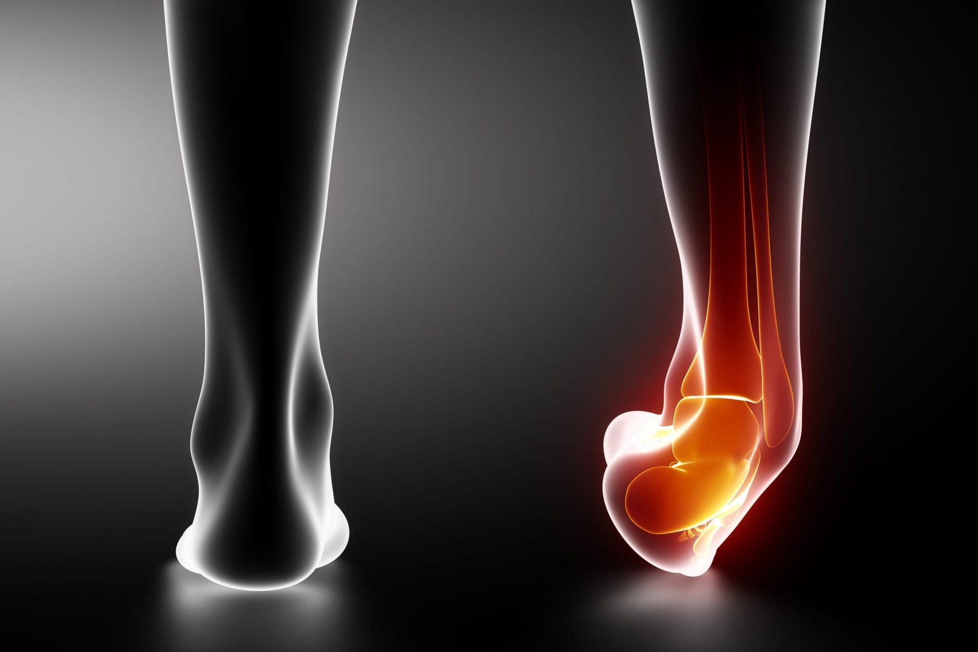
The syndesmosis is located just above the ankle where the tibia and fibula meet, providing stability to the ankle joint while allowing for its motion. A sprain, twist, rotational injury, or break to the ankle can stretch and tear the ligaments that support the syndesmosis. Syndesmotic injuries occur in up to 18 percent of all ankle sprains and 23 percent of all ankle fractures. However, the limitations of 2D imaging make a diagnosis and operative treatment of syndesmotic ankle injuries challenging.
Despite high accuracy and sensitivity, CT scans may underestimate the extent of syndesmotic lesions because of non-weight bearing conditions.
Weight bearing cone beam CT (WBCT) is an alternative imaging technique with numerous advantages, including relatively low radiation dose.
Researchers in the United States and Belgium aimed to develop a reproducible method using WBCT to quantify displacement, translation and rotation of the fibula caused by subtle syndesmostic injuries. Current methods use a single slice of a CT volume. The researchers proposed segmenting a volume out of bilateral ankle CTs superimposing the healthy ankle on the contralateral ankle to compare the deviation of the fibula to quantify the extend of the lesion.
The researchers conducted a study on eighteen patients with a unilateral syndesmotic lesion. The results were described in a study titled, “Templating of Syndesmotic Ankle Lesions by Use of 3D Analysis in Weight-bearing and Nonweightbearing CT”.
For those patients with a high ankle sprain (n = 12), bilateral imaging was performed with weight-bearing cone-beam computed tomography (CT), while non-weight-bearing CT was used for those with fracture-associated syndesmotic lesions (n = 6). To quantify the syndesmotic lesions, changes between the most lateral aspect of the lateral malleolus and the anterior and posterior tubercle in the healthy, stationary fibula were compared to those of the affected patients, using a control group of seven studies.
Deviations were calculated using defined anatomical landmarks on computer assisted design (CAD) software, rather than via manual methods.
The study found there were significant differences in the tibiofibular configuration between injured and healthy ankles.
The study concluded that
- The method was accurate in assessing subtle syndesmotic injuries.
- In the case of fracture associated with syndesmotic injury, it offered a precise description of the displacement related to the integrity of the distal tibiofibular syndesmosis.
- In a case with pronounced fibular comminution, the amount of shortening could be preoperatively calculated, facilitating reconstruction of the fibula.
Click here to read about a previous study in which cadavers were scanned via WBCT imaging in an effort to shed light on the rotational dynamics in syndesmosis.
Related Posts