When CT Is Not Enough In a recent discussion on the clinical value of weight…

Kent State Faculty and Students Research the Use of Weight Bearing CT to Effectively Assess Magnitude of Hallux Valgus Deformity
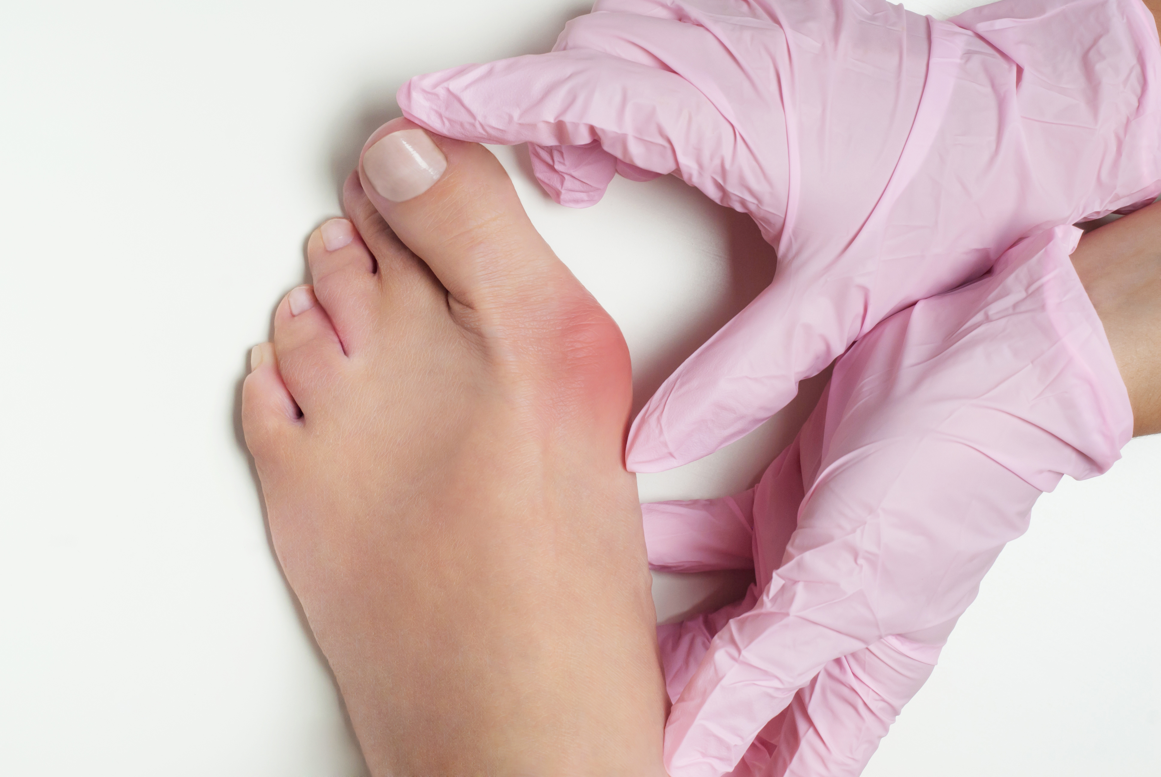
Hallux valgus is a triplane deformity. In patients with this deformity, the sesamoids displace from their normal alignment. Recent evidence suggests that, according to researchers, “the magnitude of this displacement can be determined by the coronal plane sesamoid rotation angle.” Podiatric doctors often use weight-bearing radiographs to determine the magnitude of the hallux valgus deformity. This is a crucial step in planning surgical correction. However, conventional foot radiographs have long been problematic due to:
- geometric distortion
- unreliable measurements made between different observers
- limited imaging in the coronal plane
- measurable differences between weight-bearing and non-weight-bearing images
CT scanning addresses these problems. It allows “cross-sectional imaging of the anatomical parts in all three planes of the foot without typical radiographic distortion.” In other words, doctors are able to look at slices of the foot for better data. CT scanners also allow for three-dimensional reconstruction of the foot.
The Kent State University College of Podiatic Medicine recently acquired a CurveBeam pedCAT bilateral weight-bearing CT scanner via a grant funded by the Ohio College of Podiatric Medicine (OCPM) Foundation. In the wake of this acquisition, the College of Podiatric Medicine students and faculty proposed three research projects. Each one has the potential to break new ground in the specialty. A team of KSUCPM researchers has completed the first project, which focuses on the evaluation of hallux valgus deformity in the coronal plane of the foot.
Differences in Rotation Angle Between Two Extreme Weight-Bearing Positions
Using the weight-bearing CT scanner, this study was designed to determine, according to the researchers, “the effect of different weight-bearing foot positions on the coronal plane sesamoid rotation angle as compared with standard sesamoid axial studies.” Study data demonstrated “significant differences in the rotation angle between the two extreme weight-bearing positions.” Sesamoid rotation angles, the researchers noted, were “significantly higher in the pronated foot position.” Sesamoid rotation angles from the weight-bearing CT supinated position correlated with those values.
Weight-Bearing CT Scan Determination Should Replace Forefoot Axial Studies
These results strongly suggest that “weight-bearing CT scan determination with the foot in a non-affected weight-bearing position should replace forefoot axial studies as the accepted imaging standard.”
Learn more about how Weight-Bearing CT in diagnosing Hallux Valgus patients here.
Related Posts