For Scientific Exchange Only – Not for Promotional Use. This communication includes information about an…

Iowa Researchers Use WBCT to Investigate Achilles Tendinopathy and Affects of Heel Lifts
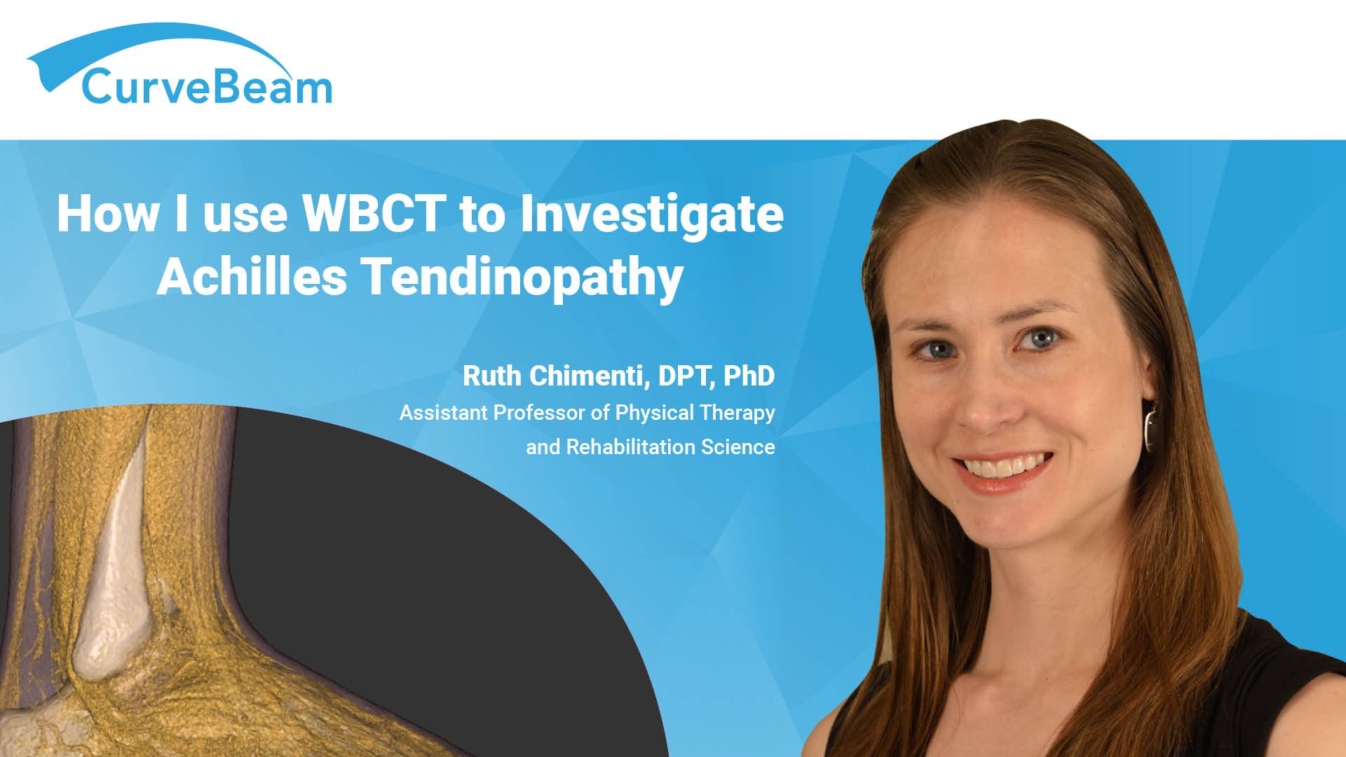
Assistant Professor of Physical Therapy and Rehabilitation Science, Ruth Chimenti, DPT, PhD, is researching the relationship between movement and musculoskeletal pain, specifically in the Achilles tendon. The University of Iowa researcher uses the HiRise system to examine localized peripheral pain mechanisms that cause tendon pain. She couples dynamic foot posture measures from a 3D motion analysis system with the HiRise’s 3D weight bearing images to determine how mechanical load affects the tendon.
One of her current studies compares wearing a heel lift to more invasive surgical procedure. HiRise weight bearing 3D imaging allows Dr. Chimenti to quantify the exact structural changes a heel lift creates in the foot.
The University of Iowa’s Orthopedic Biomechanics Laboratories’ Orthopedic Functional Imaging Research Laboratory (OFIRL) incorporates WBCT imaging into its ongoing investigations.
“Why is weight bearing CT important for the lab? I mean, it’s literally the reason for the lab to exist,” said Dr. Cesar de Cesar Netto, Director of OFIRL. “[It’s become] the gold standard for any assessment of foot and ankle pathology.”


