Fragility fractures are often the first visible sign of underlying osteoporosis but too often, they…

Distinguishing Categories of Medial Column Ligamentous Fracture
Weight bearing CT scans offer new, three-dimensional perspectives that reveal a better understanding of the biomechanics of flat foot. That was a finding on an academic poster on display at The American Orthotic and Prosthetic Association’s National Assembly in Las Vegas (Sept. 4 – 9).
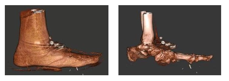
“IRD, PTTD, Posterior Tib, Flat Foot, Pronation…these are all terms that various professions use to describe some sort of medial column ligamentous breakdown,” according to the poster. “The purpose of this study is to better distinguish some types of pathomechanics of the foot. By categorizing and redefining them, it makes the description of pathology more useful for Orthotic Treatment. This was possible through the use of 3D rendering of the pedCAT machine and CurveBeam software.”
The study was conducted by Ian Engelman, M.S. CPO, and Harold Chamberlin, DPM. They looked at 3D renderings of the bony structures in flat feet, and came up with some new descriptions, as well as their possible ligamentous causes.
For example:


The study concluded that while the exterior contours of the foot suggest certain bony pathologies, the 3D renderings provided by the pedCAT would allow for orthotists to better distinguish the pathomechanics of the foot and ankle.
An unexpected result showed a moderately strong correlation (-0.68) between the vertical position of the navicular and medial displacement in a patient population with ranging degrees of medial column breakdown.


