Orthopedic decision-making depends on accurate representation of anatomy—particularly when joint alignment and bone relationships change…
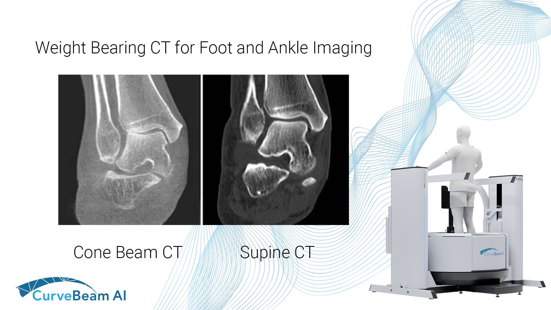
Weight Bearing CT for Foot & Ankle Imaging
Common Indications
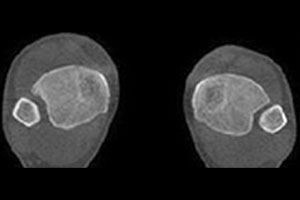
Syndesmosis
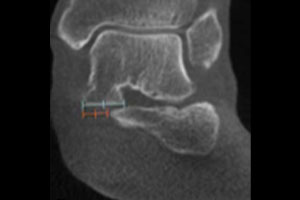
Flat Foot
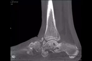
Charcot Foot
The Weight Bearing Difference
A 58-year-old patient with significant ankle pain sought a consultation with Dr. Walther. The patient had seen several orthopedic surgeons, one of whom had prescribed orthotics. However, the orthotics did not alleviate her pain. Her supine medical CT (MDCT) scan denoted only minimal arthritis. The patient could not get an MRI due to her pacemaker. Dr. Walther ordered a weight bearing CT (WBCT) scan, which revealed significantly reduced joint space. He performed a successful total ankle replacement on the patient three months later.
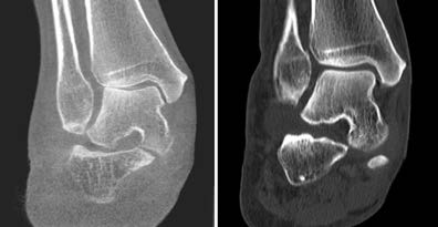
Cone Beam CT vs Supine CT

“A WBCT exam of the ankle joint and the foot allows a completely new evaluation of many deformities. It allows axis misalignments, joint instabilities, or osteoarthritis to be better classified.”
Dr. Markus Walther, MD
Foot & Ankle MD
(1) Lintz F, Bernasconi A, Ferkel EI. Can Weight-Bearing Computed Tomography Be a Game-Changer in the Assessment of Ankle Sprain and Ankle Instability? Foot Ankle Clin. 2023 Jun;28(2):283-295. doi: 10.1016/j.fcl.2023.01.003. PMID: 37137623.
(2) Hagemeijer NC, Chang SH, Abdelaziz ME, Casey JC, Waryasz GR, Guss D, DiGiovanni CW. Range of Normal and Abnormal Syndesmotic Measurements Using Weightbearing CT. Foot Ankle Int. 2019 Dec;40(12):1430-1437. doi: 10.1177/1071100719866831. Epub 2019 Aug 23. PMID: 31442094(2) de Cesar Netto C, Myerson MS, Day J, Ellis SJ, Hintermann B, Johnson JE, Sangeorzan BJ, Schon LC, Thordarson DB, Deland JT. Consensus for the Use of Weightbearing CT in the Assessment of Progressive Collapsing Foot Deformity. Foot Ankle Int. 2020 Oct;41(10):1277-1282. doi: 10.1177/1071100720950734. Epub 2020 Aug 27. PMID: 32851880.
(3) Jeng CL, Rutherford T, Hull MG, Cerrato RA, Campbell JT. Assessment of Bony Subfibular Impingement in Flatfoot Patients Using Weight-Bearing CT Scans. Foot Ankle Int. 2019 Feb;40(2):152-158. doi: 10.1177/1071100718804510. Epub 2018 Oct 8. PMID: 30293451.
(4) Wukich DK, Sung W, Wipf SA, Armstrong DG. The consequences of complacency: managing the effects of unrecognized Charcot feet. Diabet Med. 2011 Feb;28(2):195-8. doi: 10.1111/j.1464-5491.2010.03141.x. PMID:
21219429.
Related Posts