When CT Is Not Enough In a recent discussion on the clinical value of weight…
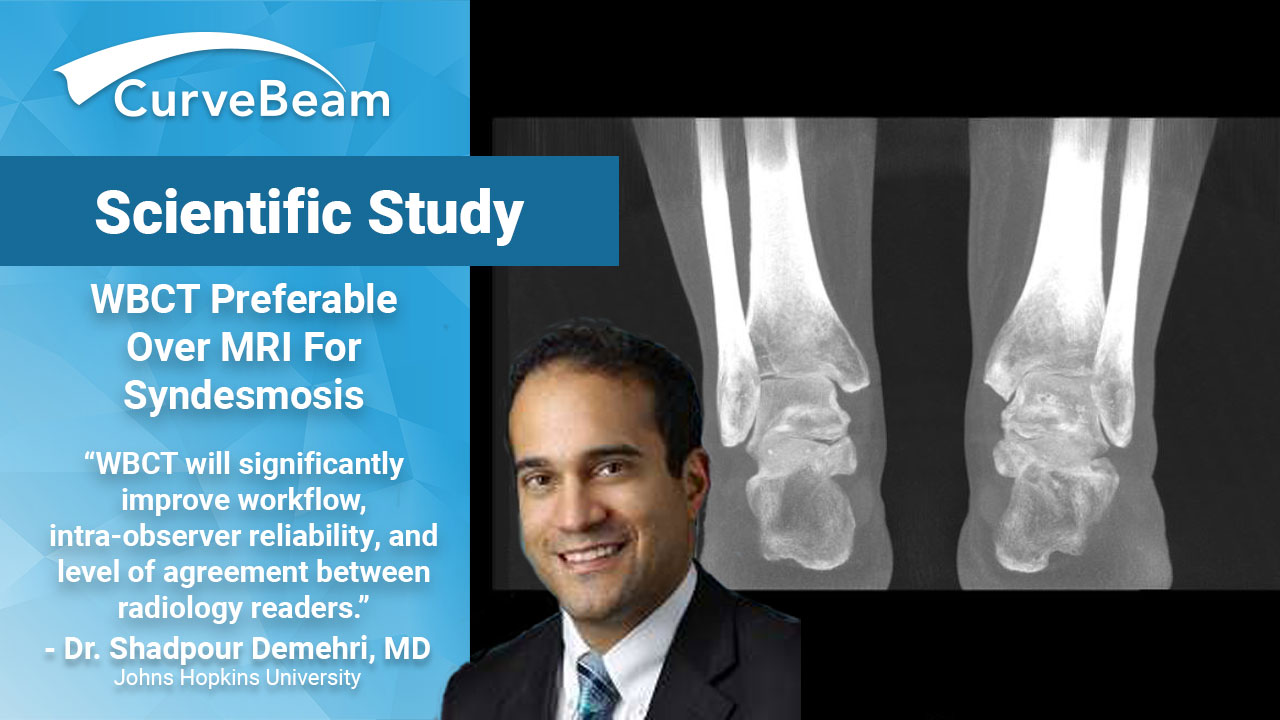
Demehri: WBCT Preferable Over MRI for Syndesmosis
 Injury to the syndesmosis often requires advanced imaging. Up until recently, the most common advanced imaging modality to evaluate this injury has been magnetic resonance imaging (MRI). Dr. Shadpour Demehri, MD, Associate Professor, Russell Morgan Department of Radiology, Johns Hopkins University, discussed the current imaging standards and the evolving role of weight bearing cone beam CT (WBCT) in a presentation he delivered to the WBCT International Study Group in July 2018.
In reviewing recent literature, Dr. Demehri explaind that MRI can reliably and accurately detect syndesmotic injury. There does not appear to be any difference between 3T and 1.5T MRI technologies, and further, there is no advantage to using contrast (MRA) when evaluating the syndesmosis. The biggest limitation of MRI studies is the lack of information regarding the significance of anatomic derangement and biomechanical instability. This limitation is where WBCT technology has emerged as a new tool for evaluation.
Dr. Demehri and his colleges conducted a study of asymptomatic patients to create an atlas of diastasis, rotation, translation, and ankle mortise measurements and range of normals. During this study, they were able to make three conclusions. First, defined syndesmotic measurements can be reliably performed on WBCT. Second, except for medial clear space, syndesmotic measurements did not vary with normal weight bearing. Finally, under physiological load, the relationship between the distal tibia and fibula remains unchanged in the presence of an intact syndesmosis.
The biggest limitation of MRI studies is the lack of information regarding the significance of anatomic derangement and biomechanical instability. This limitation is where weight bearing cone beam CT technology has emerged as a new tool for evaluation.
Considering the conclusions drawn from this initial study, Dr. Shadpour Demehri decided to further evaluate weight bearing versus non weight bearing studies in symptomatic patients. He explains that images were gathered in natural, weight bearing stance to allow for evaluation of comparative anatomy. If apparent syndesmotic injury was evident on plain x-rays or exam, those subjects were removed from the study. This allowed them to evaluate subtle injury more critically and assess the efficacy of typical patients sent for advanced imaging. The findings of this study revealed that in patients with prior ankle injuries, syndesmotic measurements using WBCT are feasible, reproducible, and had moderate to perfect level of agreement between observers. Values of syndesmotic measurements were significantly different between weight-bearing and non-weight bearing images.
As much of the radiology field continues to evolve with technology, Dr. Demehri sees great potential for the utilization of WBCT technology. He hopes to further investigate the role of automated measurements as well as automated bone segmentation and anatomical landmark localization. He believes that using these semi-automated techniques will allow measurements to be obtained using pre-defined anatomic landmarks. Ultimately, this will significantly improve workflow, intra-observer reliability, and level of agreement between radiology readers.
Watch Dr. Demehri’s full presentation here:
Related Posts