Orthopedic decision-making depends on accurate representation of anatomy—particularly when joint alignment and bone relationships change…
AOFAS Annual Meeting – CurveBeam Symposium Recap
Weight bearing CT is a vital tool for determining the cause of inexplicable pain, and also for avoiding painful surgical complications.
That was the takeaway message from a talk by Dr. Phinit Phisitkul, a clinical associate professor of orthopaedics at the University of Iowa. He shared some of his most interesting cases at a CurveBeam sponsored symposium held during the AOFAS Annual Meeting in Long Beach during the evening session.
We’ve selected three of his cases to share on this blog:
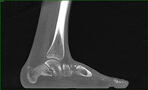
18-year-old male with Noonan Syndrome & severe flat foot: The patient presented with an unusual amount of pain that was difficult to diagnose on plain X-Ray. A weight bearing CT scan revealed he had a severe deformity – a congenital vertical talus. He also had severe impingement.


58 year-old male with ankle arthritis: The patient presented with a lot of pain in the ankle joint. A weight bearing CT scan showed a subluxation of the ankle joint and dramatic impingement of the calcaneal fibula. Interestingly, the subtalar joint was in pristine condition. Dr. Phisitkul determined the patient was a good candidate for ankle replacement and hindfoot realignment, and that his subtalar joint could be spared.

41-year-old female with Hallux Valgus: A weight bearing CT scan revealed a bone spur on the patient’s first metatarsal head. If the doctor had done a normal release, the spur may have ended up pinching the sesamoid. Instead, he performed a lateral release and excised the bone spur.

Related Posts