We are thrilled to announce an esteemed panel of speakers who are at the forefront…
Webinar Summary: Advanced CT Imaging in Foot and Ankle Surgical Considerations
We all know that X-rays and MRIs aren’t the be-all, end-all of diagnostic imagery.
Try determining the frontal plane rotation of the sesamoid and first metatarsal with an X-ray. It isn’t possible. Correction of the hallux valgus rotation in bunion surgery depends largely on the repositioning of the sesamoidal apparatus which is impossible to assess without an axial view, and X-rays fall completely short when it comes to assessing these relational details from the vantage point of a single plane.
Such was the topic of discussion during the latest Curvebeam webinar led by Dr. Bob Baravarian, Director of University Foot and Ankle Institute in Southern California. He explained how weight-bearing 3-dimensional CT scans are changing the game of podiatric diagnostics.
Throughout the webinar, titled “Advanced CT Imaging in Foot and Ankle Surgical Considerations,” Dr. Baravarian offered a very straightforward presentation of the often-not-so-straightforward complexities of foot and ankle deformities and how advanced imaging technology can improve both surgical planning and surgical outcomes. He cited the example of hallux valgus among many others as “impossible to imagine treating without 3-D imaging technology” these days – given the results he’s seen with his patients and scope 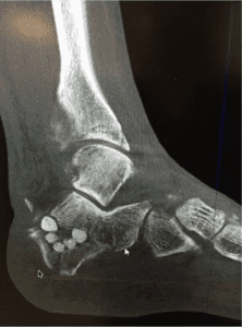 of its applications.
Planning the frontal plane correction of first metatarsal.
“CT scans are very helpful in planning your frontal plane deformity correction of the first metatarsal to get an anatomic position which is really underestimated,” said Dr. Baravarian.
Multi-plane imaging now allows physicians to correct the frontal plane deformity of the metatarsal while simultaneously realigning the sesamoid. The capability is “critical,” said Dr. Baravarian, “for proper outcome with bunion corrections whether you’re doing a LAP or any kind of osteotomy.”
Identifying the cause of hypermobility of the first ray
When you look at a patient who has a significant flat foot deformity and a significant bunion deformity with some level of hyper-mobility of the first ray, 3D CT imaging allows you to locate the exact area in need of correction.
“In patients who have PTTD or even a pediatric flat foot case we really need to decide which planes of correction make the best sense,” said Dr. Baravarian. “If I correct my first metatarsal, is my hind foot going to realign or is there an outstanding deformity that needs correction?”
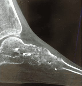 Determining the level of arthritis in hallux rigidus and limitus cases.
“I’m constantly surprised when I go into surgery and I plan a cheilectomy and I open up a joint and there’s significantly more arthritic changes or some kind of osteochondral legion that I couldn’t really see on the X-ray. Or, I plan to do an osteotomy and I go in and the level of arthritis is not as bad as expected,” said Dr. Baravarian.
It’s very difficult to determine the level of arthritis in a hallux rigidus or limitus case based on a radiograph alone. While MRIs are an excellent option for soft tissue imaging and diagnostic ultrasounds still provide accurate, real-time guidance for proper injections –neither of these methods makes sense for treating anatomic alignment and assessing structural deformities.
“With a 3D CT I’m able to look a little more in-depth into the joint and make a better decision prior to surgery.”
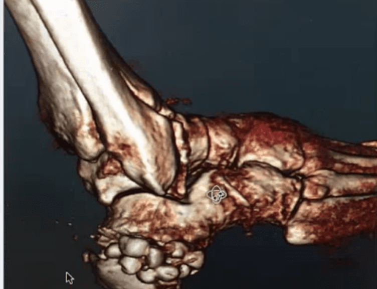
Identifying major deformities in complex fractures.
Weight-bearing imaging systems allow you to place a foot in its anatomic position and adjust its deformity to see what level of correction you can get across multiple planes.
It “doesn’t makes sense,” according to Dr. Baravarian, “to plan the correction of a complex fracture without a sense of what’s going on inside the foot and ankle.”
Our traditional method of two-dimensional x-rays doesn’t provide the highest level of certainty that doctors need in order to administer the best care possible to their patients where deformity planning is essential for proper surgical outcomes.
Now, with the help of tools such as the pedCAT, a compact 3D weight-bearing CT imaging system, podiatrists have everything they need to create comprehensive treatment plans and more effective surgeries. Better outcomes. Less risk. And patients back on their feet faster than they ever expected.
You can access the entire webinar here. A FOOT Innovate membership is required to access the content. Membership is complimentary for foot & ankle specialists.

