We are thrilled to announce an esteemed panel of speakers who are at the forefront…
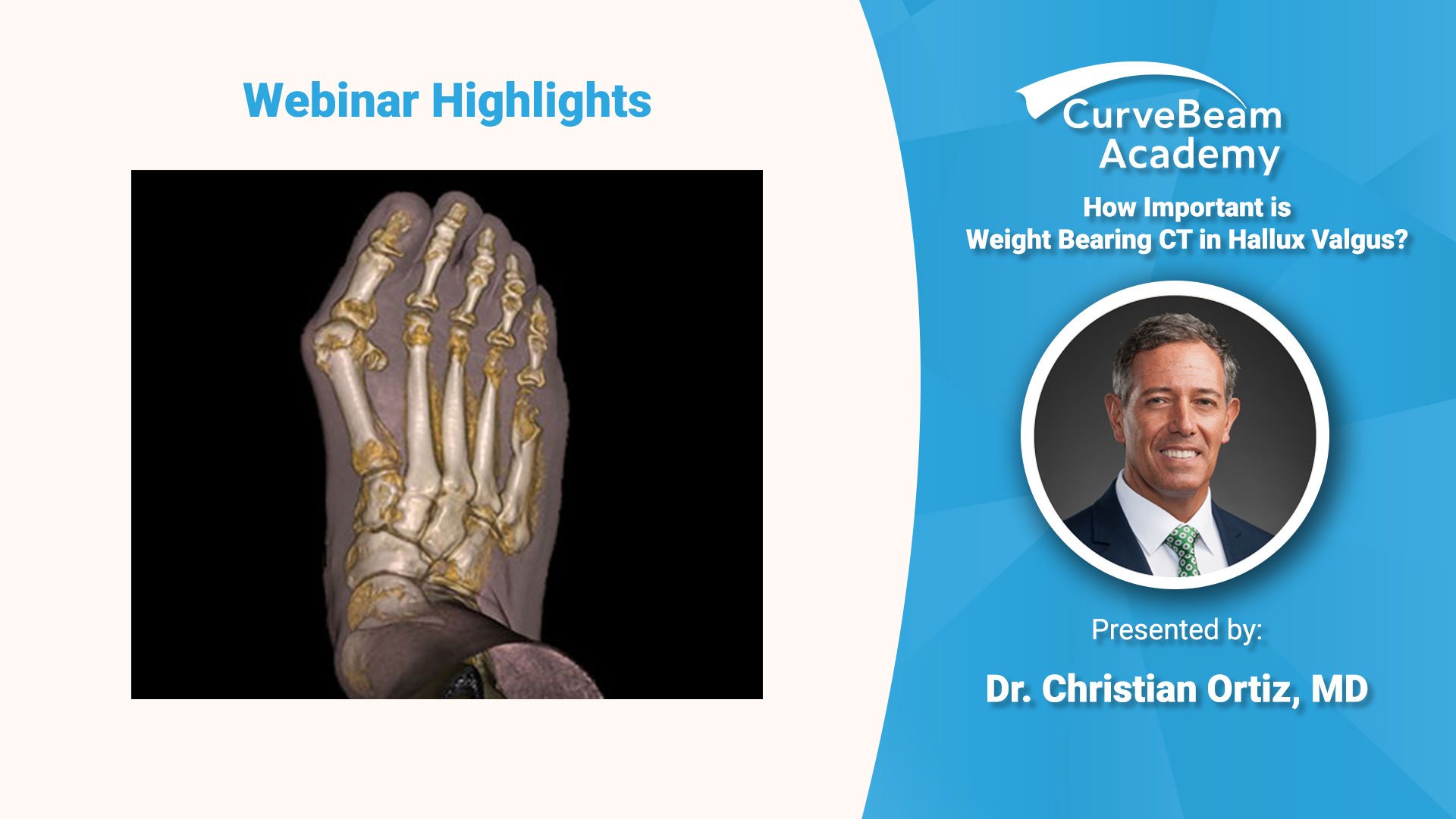
WBCT & Rotation in Complex Hallux Valgus Cases
Dr. Cristian Ortiz, a foot and ankle orthopedic surgeon at Clinica Universided de los Andes, discussed what role weight bearing CT (WBCT) imaging plays in the assessment of hallux valgus (HV) as part of the CurveBeam Academy: Focus on Foot & Ankle webinar series.
Dr. Ortiz emphasized that when it comes to HV, rotation exists, but is often missed, leading to deformity recurrences. Click below to watch a condensed version of the webinar and get the highlights in less than 10 minutes.
To access the full length webinar, please click here.
“Weight bearing CT is absolutely necessary and from my point of view it replaces X-Rays, ” Dr. Ortiz said. “Every patient who is a candidate for hallux valgus surgery in my hospital gets a WBCT pre-op. I still have recurrences. I still have failures. But I think my failures have been lessened, now that I have this very useful tool available for my decision making process.”
Dr. Ortiz reviewed a recent paper published by Dr. Cesar de Cesar Netto and Dr. Martinus Richter, in which they used WBCT to analyze HV deformities. They determined HV often includes hypermobility not only the 1st TMT, but the whole 1st ray. Dr. Ortiz commented that for some hallux valgus cases, WBCT could help surgeons plan in advance what part of the medial column needs to be fixed. Netto & Richter concluded HV is obviously a measurable triplanar deformity, but we are still missing key information to determine normal versus abnormal ranges.
Ortiz and his colleagues have scanned more than 400 HV cases, and are documenting the degree of sagittal instability and rotation in every plane.
Dr. Ortiz presented his decision table for hallux valgus surgeries.
– For deformities where the angle to be corrected is less than 5 degrees, he will perform a Chevron osteotomy.
– For deformities where the angle correction required is 5 – 10 degrees, a Scarf osteotomy is his go-to method. He said a de-rotation osteotomy could be added, however, he has observed that “if you correct alignment in one plane, rotation takes care of itself.”
– For cases where the angle correction is more than 10 degrees, options are a proximal osteotomy or Lapidus bunionectomy. It is important to keep in mind that you need to achieve rotation at the right location to have proper correction, he stressed.
Ortiz’s clinic is currently working on a study where they obtain a WBCT scan of post-op as well to quantitatively measure what corrections they are achieving. Dr. Ortiz said the results will influence their decision making process in the future.
To access the full length webinar, please click here.
Related Posts