AI-Driven Segmentation on Weight Bearing CT Introduction Accurate segmentation is critical for orthopedic workflows, including…
Scientific Literature: The New Standard? A Comparison of 2D and 3D Measurements of the Foot & Ankle Via Weight Bearing CT Imaging
A group of radiologists from the Netherlands and Italy are asking if 3D angle measurements taken on weight bearing CT scans should be the new standard for foot & ankle evaluation.
Automated 3D analyses of the foot & ankle using weight bearing cone beam CT (CBCT) are more reproducible and precise compared to 2D analyses, according to a 2021 study published in the European Journal of Radiology. In addition, 3D evaluation better demonstrates differences in bone configurations between weight bearing and non-weight bearing conditions, which may be of value to demonstrate pathology.
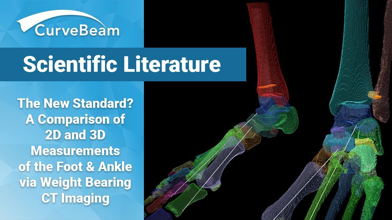
The researchers examined 20 healthy volunteers who each had weight bearing and non-weight bearing CBCT scans performed on both feet. Clinically relevant height and angle measurements were performed in both 2D and 3D. The 2D measurements were derived from Digitally Reconstructed Radiographs (DRR)created by HOROS software. CurveBeam provides DRRs via it’s Insta-X feature in its CubeVue visualization software. Angles between bones were then defined manually by drawing anatomical axis lines through the center of the body structures in 2D sagittal and/or axial views of the foot.
3D measurements were obtained using software which automatically composes anatomical long-axes of the bone structures based on their inertial axes. The 3D datasets were first manually segmented, a process which took about 6 hours per research subject.
While intra and inter observer agreement was good on the 2D measurements, the coefficient of variation in 3D parameters was generally lower. The lower intraclass correlation coefficients (ICC) on 2D measurements were likely due to over projection of 1st metatarsal and difficulty in determining the long axis of bones such as the calcaneus and talus.
No significant differences were found between the left and right foot in weight bearing or non-weight bearing position in either 2D or 3D measurements, an indication that “the contralateral side could serve as a reference for corrective surgery,” according to the study.
Today’s current standard of care for assessing pathological foot morphology and severe deformities is often weight bearing radiographs [1-3]. However, this two-dimensional method often fails to accurately represent complex foot structures, and factors such as over-projection of the bones can affect the reliability of analyses [1].
When using a CBCT system, 3D images of the lower extremities in a weight bearing or non-weight bearing natural standing position can be acquired, allowing for images to reveal issues that may not have normally been seen in 2D scans [4-8]. Studies show that 3D weight bearing CT is more useful than 2D imaging, since 3D data allows for better multi-planar insights of the pathology [3,9-11].
Researchers suggested that future studies should investigate the possible differences between male and females, the value of comparing geometric parameters between weight bearing and non weight bearing images, and comparing them with the healthy contralateral side in a group of patients with unilateral disorders.
[1] Z.B. Cheung, M.S. Myerson, J. Tracey, E. Vulcano, Weightbearing CT scan assessment of foot alignment in patients with hallux rigidus, Foot Ankle Int. 39 (2018) 67–74, https://doi.org/10.1177/1071100717732549.
[2] J.Z. Zhang, A. Bernasconi, S. Zhang, 3D biometrics for hindfoot alignment using weightbearing computed tomography, Foot Ankle Int. 60 (2019) 720–726, https:// doi.org/10.1177/1071100719835492.
[3] A. Burssens, J.P.M. Peiffer, R.M.T. Lenaerts, W. Isg, G.V.J. Victor, Reliability and correlation analysis of computed methods to convert conventional 2D radiological hindfoot measurements to a 3D setting using weightbearing CT, Int. J. Comput. Assist. Radiol. Surg. 13 (2018) 1999–2008, https://doi.org/10.1007/s11548-018- 1727-5.
[4] A. Barg, T. Bailey, M. Richter, C. de Cesar Netto, F. Lintz, A. Burssens, et al., Weightbearing computed tomography of the foot and ankle: emerging technology topical review, Foot Ankle Int. 39 (2018) 376–386, https://doi.org/10.1177/ 1071100717740330.
[5] J.A. Carrino, A. Al Muhit, W. Zbijewski, G.K. Thawait, J.W. Stayman, N. Packard, et al., Dedicated cone-beam CT system for extremity imaging, Radiology 270 (2014) 816–824, https://doi.org/10.1148/radiol.13130225.
[6] E.K.J. Tuominen, J. Kankare, S.K. Koskinen, K.T. Mattila, Weight-bearing CT imaging of the lower extremity, Am. J. Roentgenol. 200 (2013) 146–148, https:// doi.org/10.2214/AJR.12.8481.
[7] T. Kimura, M. Kubota, T. Taguchi, N. Suzuki, A. Hattori, K. Marumo, Evaluation of first-ray mobility in patients with hallux valgus using weight-bearing CT and a 3-D analysis system a comparison with normal feet, J. Bone Jt. Surg. Am. 99 (2017) 247–255, https://doi.org/10.2106/JBJS.16.00542.
[8] A. Hirschmann, C.W.A. Pfirrmann, G. Klammer, N. Espinosa, F.M. Buck, Upright Cone CT of the hindfoot: comparison of the non-weight-bearing with the upright weight-bearing position, Eur. Radiol. 24 (2014) 553–558, https://doi.org/ 10.1007/s00330-013-3028-2.
[9] C. de Cesar Netto, L.C. Schon, G.K. Thawait, L.F. da Fonseca, A. Chinanuvathana, W.B. Zbijewski, et al., Flexible adult acquired flatfoot deformity, J. Bone Jt. Surg. Am. 99 (2017) 1–12, https://doi.org/10.2106/JBJS.16.01366.
[10] A. Burssens, H. Vermue, A. Barg, N. Krahenbühl, ¨ J. Victor, K. Buedts, Templating of syndesmotic ankle lesions by use of 3D analysis in Weightbearing and nonweightbearing CT, Foot Ankle Int. 39 (2018) 1487–1496, https://doi.org/ 10.1177/1071100718791834.
[11] B. Campbell, M.C. Miller, L. Williams, S.F. Conti, Pilot study of a 3-Dimensional method for analysis of pronation of the first metatarsal of hallux Valgus patients, Foot Ankle Int. 39 (2018) 1449–1456, https://doi.org/10.1177/ 1071100718793391.
Related Posts


