WBCT on the Program Thursday, January 22 11:10 am – 12:30 pm Symposium 2: Diagnostic…
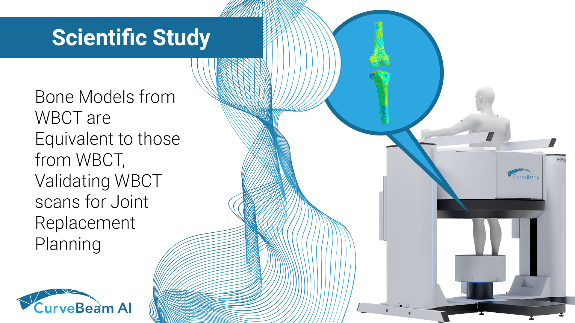
New Study Confirms WBCT Delivers Equivalent 3D Bone Models to MDCT for Joint Replacement Planning
A groundbreaking study published in Scientific Reports confirms that weight-bearing cone beam CT (WBCT) produces 3D bone models of the knee that are statistically equivalent to those generated from multi-detector CT (MDCT). This reinforces WBCT’s readiness for integration into surgical planning workflows—especially in robotic-assisted total knee arthroplasty (TKA).
The study, titled “Equivalence assessment of weight bearing cone beam CT and multidetector CT through 3D Knee bone modelling,” was conducted by Xiaoxu Li, PhD (Monash University) and researchers from CurveBeam AI, Tennessee Orthopaedic Alliance, and the Hip and Knee Clinic in Belgium. Their objective was clear: assess whether WBCT provides the same geometric fidelity needed for clinical applications as MDCT, the current standard.
Study Highlights:
- Data Source: 3D models were generated from both WBCT and MDCT scans of four patients and ten cadaver knees.
- Evaluation Across All Four Knee Bones: Distal femur, proximal tibia, proximal fibula, and patella.
- Manual Segmentation: Models were manually segmented using a multi-review process to ensure clinical accuracy.
- Mesh-Based Comparison: Rigid registration and vertex-level surface distance analysis allowed precise measurement of shape differences between modalities.
- Submillimeter Variations: Differences between WBCT and MDCT models were consistently within a ±0.15 mm equivalence margin.
Key Findings:
- Statistically Equivalent Models: Using the Two One-Sided Tests (TOST) approach, all 3D bone models from WBCT were found to be statistically equivalent to those from MDCT.
- Consistent Across Scanners and Protocols: Despite different acquisition protocols, scanners, and reconstruction settings, the 3D model quality remained robust and clinically consistent.
- High Fidelity in Bilateral Scans: WBCT retained accuracy even when both knees were scanned simultaneously, a common clinical need.
- Visualization Confirms Minor Discrepancies: Color-coded mesh distance maps showed mostly light tones across bone surfaces, signifying minimal localized differences.
Why This Matters for Surgical Planning Software
Robotic and navigated orthopedic systems routinely accept CT scans from various vendors and settings. The consistency demonstrated in this study suggests that 3D bone model generation is resilient to modality and scan parameters. WBCT’s ability to capture images under physiological load offers additional insight that static imaging can’t provide—without sacrificing geometric precision.
With an upright posture imaging, and now proven 3D model equivalence to MDCT, WBCT is an ideal solution for practices aiming to optimize preoperative workflows in joint replacement.
Li, X., Ivie, C., Overschelde, P.V. et al. Equivalence assessment of weight bearing cone beam CT and multidetector CT through 3D Knee bone modelling. Sci Rep 15, 20974 (2025). https://doi.org/10.1038/s41598-025-06626-1
Related Posts