AI-Driven Segmentation on Weight Bearing CT Introduction Accurate segmentation is critical for orthopedic workflows, including…
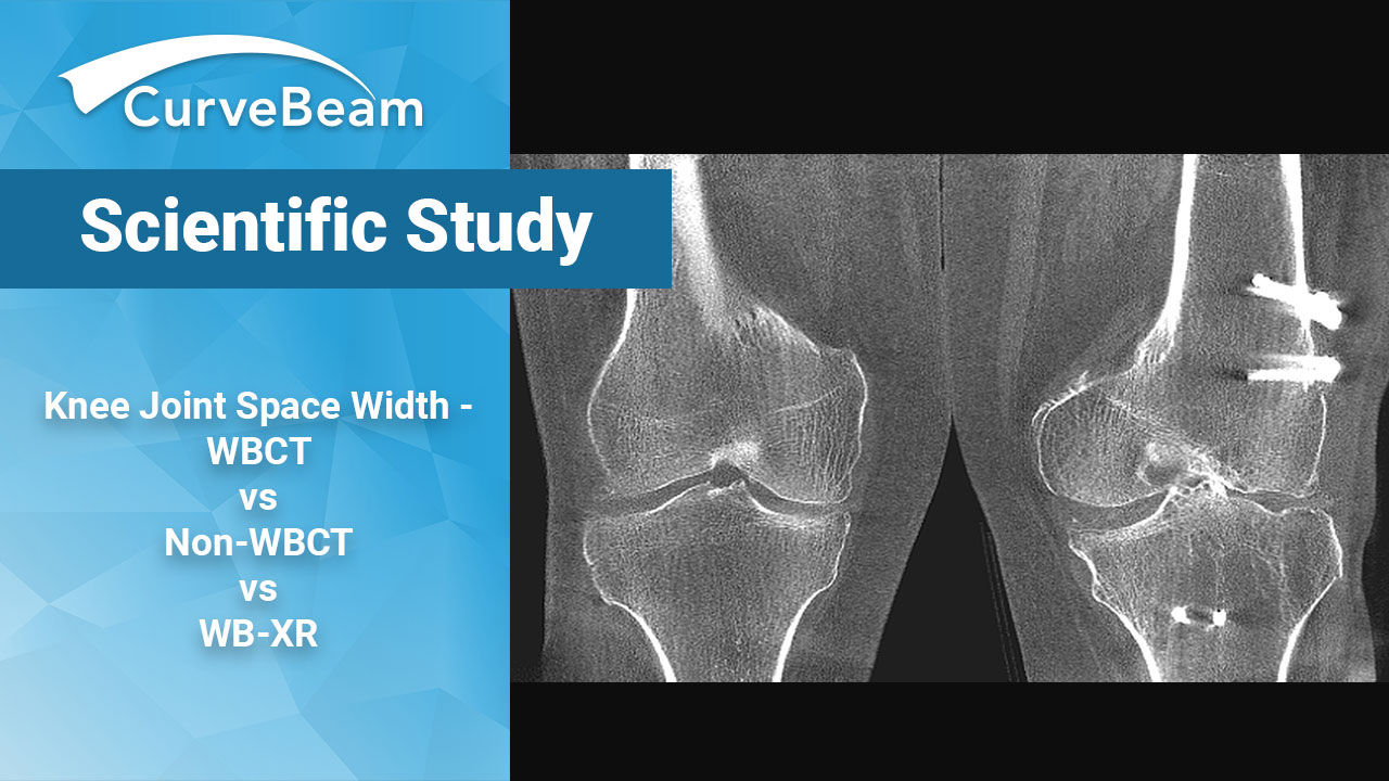
Knee Joint Space Width – WBCT vs Non-WBCT vs WB-XR
3D Quantification of Knee Joint Space Width with WBCT, Non-WBCT & WBXR: A Comparison
Key Points:
- On WBCT, 25% of the femorotibial compartments showed bone-on bone apposition, which was significantly higher when compared to NWB-CT and WB-XR.
- For patients with varus or valgus knee alignment, significant differences in joint space width (JSW) were detected between WBCT and non-WBCT scans.
- The minimal JSW on WB-XR were significantly wider compared to WBCT and NWB-CT.
Joint space narrowing is a classic characteristic of knee joint degeneration. However, standard non-weight bearing CT (NWB-CT) examinations of the extent of joint space width often misdiagnose or give a false impression of the preserved articular cartilage. The introduction of weight bearing CT (WBCT) has heralded a true view of the joints under physiological conditions.
A recent study from the Balgrist University Hospital in Zurich, Switzerland showed that WBCT demonstrates significantly more bone-on-bone appositions and shows detailed quantification of the femorotibial joint space and the effect of knee alignment on joint space width (JSW), an issue often underestimated or even undetected with NWB-CT and weight bearing X-Ray (WB-XR). The study by Dr. Benjamin Fritz et al was published in the December issue of Osteoarthritis and Cartilage.
Materials and Methods
Twenty-six participants met the inclusion criteria for the study. All exams were acquired during a previously conducted prospective study.
NWB-CT scans were performed in a supine position with fully extended knee joints on a 64-slice CT scanner (axial slice thickness was between 0.67mm and 0.9mm with an overlap of 50). WBCT scans were performed in a standing position with the knee joint in full extension in a cone beam CT scanner (axial slice thickness was 0.4 mm with no overlap). WB-XR scans were performed in a standing position with the knee joint in full extension in anteroposterior direction with a film-to-focus distance of 1.5m. The direction of the central X-Ray beam was strictly horizontal and centered on the patellar apex.
Medial and lateral femorotibial joint spaces were calculated on NWB-CT and WBCT images.
Software was used to assess the minimal distance of any point of the segmented subchondral bone surfaces to the distal femur. Color-Coded maps that indicated the minimal distance of any point on the medial and lateral subchondral bone surface to the femur were then generated.
Grading of osteoarthritis of the knee joint was assessed in all participants on anteroposterior, standing, WB-XR in full extension according to the Kellgren and Lawrence (KL) classification system for osteoarthritis. Bone-on-bone apposition was defined as a distance of less than 0.4 mm between the femoral condyle and the tibial plateau due to the voxel size of WBCT and visual inspection and correlation. 0.4 mm is considered to represent the detection limit of this 3D approach for quantitative JSW analysis.
Twenty-two of the participants underwent additional WB-XR, which were evaluated for mechanical leg axis determination. The mechanical leg axis was defined as the angle between the line from the center of the femoral head to the center of the intercondylar notch and the line from the center of the tibial eminence to the tibiotalar joint center. Varus and valgus knee deformities were defined as an angle of more than 3 degrees of the mechanical leg axis in its respective direction.
Results
Significant differences existed of the average medial and lateral JSW between WBCT and NWB-CT. The minimal JSW on WB-XR were significantly wider compared to WBCT and NWB-CT, but not significantly different between WBCT and NWB-CT. Significant differences between WBCT and NWB-CT existed in participants with varus knee alignment for the average and the minimal medial JSW and for participants with valgus alignment for the average lateral JSW. On WBCT, 25% of the femorotibial compartments showed bone-on bone apposition, which was significantly higher when compared to NWB-CT and WB-XR.
Conclusion
WBCT, and the 3D aspects of the scans, allows detailed quantification of the femorotibial joint space and the effect of knee alignment on JSW. WBCT demonstrates significantly more bone-on-bone appositions, which are underestimated or even undetectable on NWB-CT and WB-XR.
To read the full article click here.
Related Posts


