Read the article.

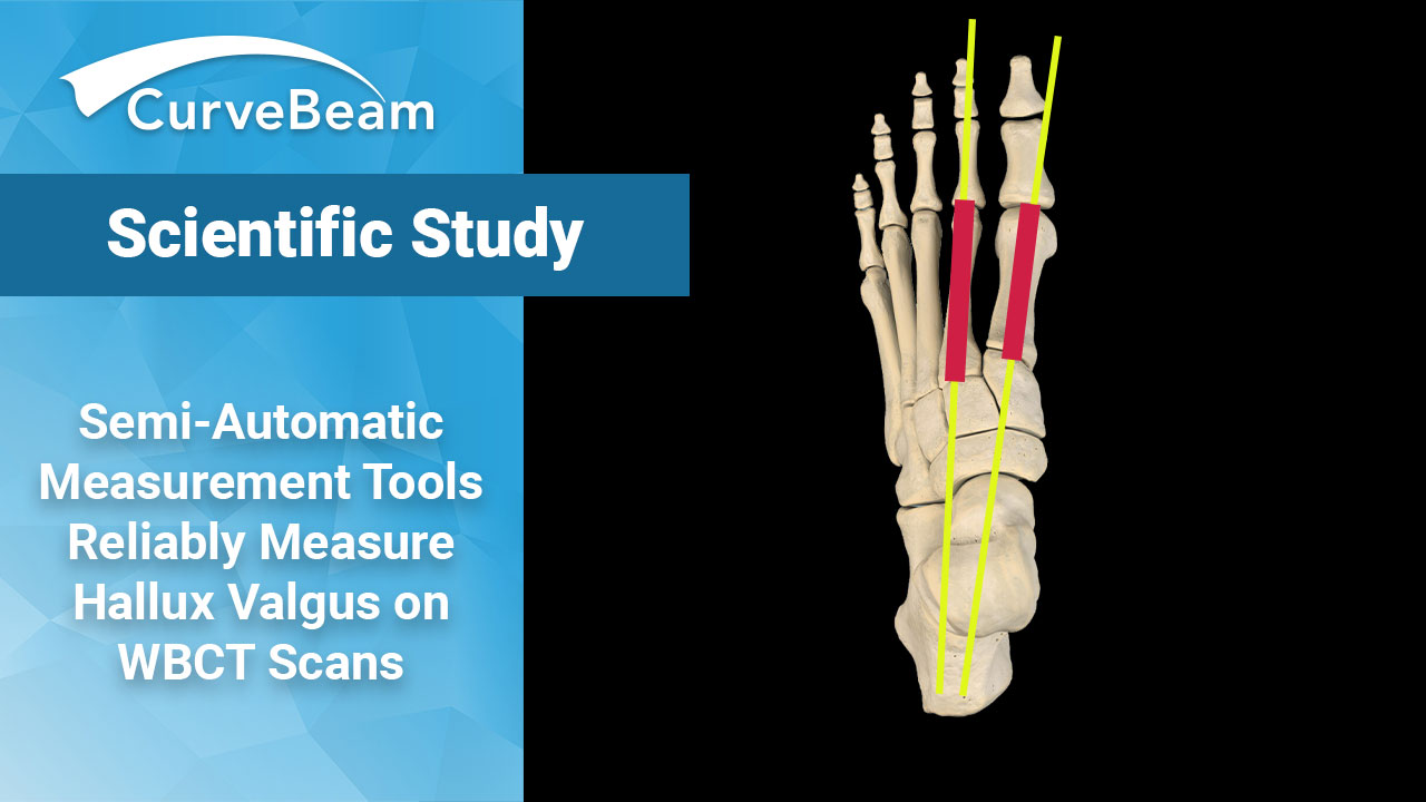
Comparison Between Weight Bearing CT Semi-Automatic and Manual Measurements in Hallux Valgus
Key Points
- Semi-automatic measurements made on WBCT scans are reproducible and comparable to measurements performed manually.
- Software designed for WBCT help differentiate pathological from non-pathological conditions.
- Semi-automatic software tools can help orthopedic surgeons obtain more accurate diagnosis with WBCT and lead to a treatment plan based on more reliable data.
The advent of weight bearing CT (WBCT) has led to the foot & ankle specialty to discuss whether it is time to update the standard of care for diagnosing, planning treatment for, and assessing outcomes for Hallux Valgus (HV). A recent study shows new software tools designed specifically for making semi-automated measurements on WBCT scans are reliable and reproducible when it comes to making common HV measurements, making the transition from traditional weight bearing X-Rays to WBCT that much smoother.
Dr. Keplar Alencar Mendes de Carvalho et al. out of the Carver College of Medicine, Department of Orthopedics and Rehabilitation, University of Iowa, Iowa City, IA, found semi-automated software tools could not only measure, but also differentiated pathological from non-pathological conditions.
Materials and Methods
The study compared WBCT scans of 26 HV feet with a control group was also assessed consisting of 19 feet.
WBCT scans were performed with CurveBeam’s pedCAT. Using CubeVue CurveBeam’s custom visualization software, hallux valgus angle (HVA), Intermetatarsal angle (IMA) and Interphalangeal angle (IPA) were recorded in the axial plane; the metatarsal and phalanx axes were established in the axial plane; and angular measurements were performed using the Cobb method.
The same surgeon investigators then performed the semi-automatic 3D measurements in a third-party software solution. The longitudinal axis estimate was generated automatically for each patient-specific metatarsal and phalanx model by first finding the center of the specific bone in proximal to distal orientation and by analyzing its cross sections at different locations. In sequence, the software selected the straight-line representative for the center of the bone by applying robust line-fitting routines. The software then automatically registered a mathematical model of the foot and ankle on the image and computed the location of measurement landmarks and longitudinal axes of bones of interest.
Results
Reliabilities utilizing interclass correlations (ICC) were over 0.80 for WBCT manual measurements and WBCT semi-automatic readings, intraobserver reliability proved to be excellent in both cases, however, the intraobserver reliability for semi-automatic HVA, IMA, and IPA was superior to manual measurements. WBCT measurements assessed by ICC (interobserver agreement and consistency) were excellent in both cases, though HVA, IMA, and IPA semi-automatic measurements showed the highest values when compared to manual measurements.
According to Pearson’s coefficient, there is a strong positive linear correlation for HVA, IMA, and IPA when comparing semi-automatic and manual measuring on patients with HV. Utilizing Bland-Altman plots, excellent agreement between semi-automatic and manual measurements for HVA, IMA and IPA was found. The graph showed that the mean difference between measurements for HVA was 3.11 degrees, with a 95% confidence interval of (1.26 – 4.97), for IMA it was 1.09 degrees, with CI (0.52 – 1.65) and for IPA it was 0.93 degrees, with CI (0.06 – 1.93) indicating a high correlation between the parameters calculated from both measurement methods and a strong agreement between the readers and the software.
Conclusion
The study concluded that semi-automatic measurements are reproducible and comparable to measurements performed manually. The software could differentiate pathological from non-pathological conditions when subjected to semi-automatic measurements.
Researchers noted that the technology is still under development and is currently restricted to few research centers, resulting in the need for future studies to discover how WBCT can integrate into daily clinical practice and speed up the decision-making process.
Researchers further noted that fully automated segmentation would represent a crucial advance in evaluating HV and other complex foot deformities. CurveBeam’s Autometrics platform, which is based on deep machine learning, will provide fully automated HV measurements. Autometrics is investigational only and is not available for sale.
To read the full study click here.


