Read the article.

Bio-Screws Could Eliminate Need for MAR Protocols in Post-Op Imaging
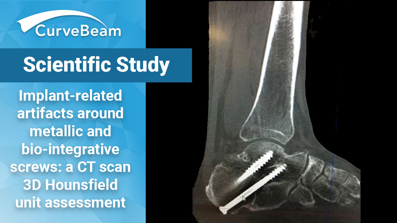
 Bio-integrative (BI) cannulated screws may be a preferred alternative to metallic implants to decrease implant-related artifacts (IRA) as they may remove the need for special CT metal artifact reduction (MAR) algorithms, according to a recent study by Dr. Christian VandeLune et al, Department of Orthopedics and Rehabilitation at University of Iowa in Iowa City.
Dr. Tutku Tazegul, one of the authors on the study (pictured left), was recently awarded for “Research Excellence in Orthopedics” by the University of Iowa Carver School of Medicine for this study.
Assessing Bone Healing – Challenges
Assessment of bone healing in osteotomies, fractures, and fusions continue to prove challenging for orthopedic surgeons, especially post-operatively. IRA can hinder the assessment of union and healing. Traditionally, both clinical and radiographic imaging are the methods used to assess any pain, site mobility issues, implant failure, and bone bridging to decide if the union was successful. CT imaging is widely regarded as the gold-standard method when assessing bone healing due to its fine trabecular detail, but there is limited literature comparing the severity of IRA when using metallic or BI implants.
Metallic vs. BI Cannulated Screws
Dr. VandeLune’s objective was to compare the degree of IRA when using metallic vs BI cannulated screws via Hounsfield unit (HU) measurements on CT. His team hypothesized that BI implants would demonstrate a significantly decreased IRA around the inserted screws.
Study Design
The study used two fresh-frozen cadaveric lower legs that had no deformities. Once thawed, a fellow-ship trained foot and ankle orthopedic surgeon with more than 10 years of experience, performed a medial displacement calcaneal osteotomy on both specimens using a 5cm traditional oblique lateral heel approach. One specimen received two 4.0mm standard cannulated metallic (titanium) screws (Wright Medical R), while the other received two 4.0mm cannulated BI fiber screws (OSSIO R).
Both cadavers were scanned post-operatively on a CurveBeam HiRise weight bearing CT system at the University of Iowa’s Orthopedic Functional Imaging Research Laboratory (OFIRL).
HU distribution was analyzed using CubeVue, CurveBeam’s custom visualization software.
The overall HU distribution within a 3D cube of 30mm edge length was assessed. Additionally, within the cube, 4 linear projections were selected to sample the entire set of HU values along that line. Line 1 was placed in close proximity to the screw (within 1cm), Line 2 was placed directly over the screws, Line 3 was placed inside the screw cannula, and Line 4 was placed dorsal to the implant by 1cm. The HU values on these lines were measured across the entire length of the line, Additionally, an 8mm segment of each line as it crossed the osteotomy was selected.
Study Results
Results showed that the metallic screws had significantly higher HU values for lines 1, 2, and 3 when compared to the BI screw specimen. Values were significantly increased when considering both the entire line inside the 3D cube as well as the selected 8mm line segment across the calcaneal osteotomy. When considering Line 1 (close to the screw), the metallic implant had higher HU values (entire, 7.26; selected, -5) when compared to BI (entire, -159; selected, -249). For Line 2 (over the top of the screw), the metallic implant was found to have higher HU values (entire, 4.84; selected, 6286) and the BI had lower values (entire, 108; selected, 151.2). Line 3 (inside the cannula) was also found to have increased HU values for the metallic implant (entire, 1664; selected, 198.7) compared to the BI one (entire, 144; selected, -277.7). However, across Line 4 (1cm away from the implant), HU values were significantly decreased around the metallic implant (entire, -49: selected, -110) compared to the BI implant (entire, 178.5; selected, 221).
Researchers concluded that BI implants significantly reduced HU values in 3 different planes across a calcaneal osteotomy. Interestingly, HU values 1cm away from the metallic screw (Line 4) were lower than those around the BI screw, potentially due to a beam hardening artifact.
Potential Impact on Imaging
BI implants could possibly remove the need for special CT protocols/software that utilize metal artifact reduction (MAR) algorithms. This could lead to improved scan quality around the implant, while saving resources on imaging and processing. Researchers stated that more studies are needed to compare IRA between metallic implants and BI implants.
To read the full article click here.


