Orthopedic decision-making depends on accurate representation of anatomy—particularly when joint alignment and bone relationships change…

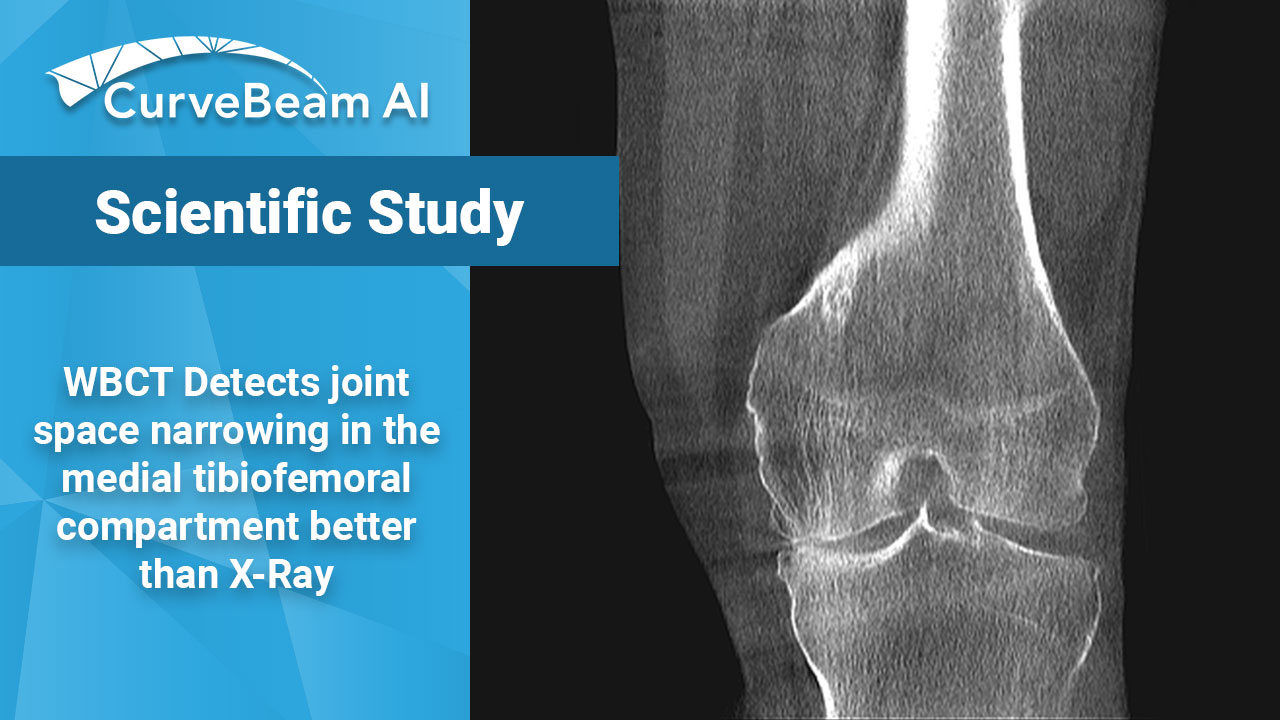
WBCT Better Detects Changes in Knee Joint Space Width than X-Ray in OA Patients
Key Points:
- Weight Bearing (WB) radiographs are limited by 1) their 2D nature and 2) knee positioning. X-Ray (XR) beam angle differs by person and can be unreliable.
- Weight Bearing CT (WBCT) imaging provides three dimensional (3D) images of the knee compartment that could allow for earlier detection of osteoarthritis (OA).
- WBCT allows for additional measurements to aid in a more reliable diagnosis of OA.
OA is the most prevalent form of arthritis and one of the most common causes of disability in the US; the knee being the most affected weight bearing joint. The current standard-of-care is to measure joint space width (JSW) on plain radiographs to diagnose and visualize the structural changes related to knee OA.
Two-dimensional (2D) JSW is typically measured as the distance between projected femoral and tibial bone margins on WB coronal knee radiographs. There are, however, limitations in this approach. Optimal knee positioning and X-Ray beam angle differs by person and can be unreliable with WB radiographs. Additionally, tibial and femoral point selection can be challenging, as sagittal tibiofemoral alignment cannot be clearly distinguished on the coronal plane.
WBCT imaging, such as CurveBeam AI’s HiRise, addresses these limitations by providing 3D images that can be obtained in a standardized, reproducible, standing position. WBCT imaging could result in earlier detection, mitigating the long-term effects of OA.
Dr. N.A. Segal et al. out of the University of Kansas, Lawrence, Kansas, aimed to compare measurements of JSW on WBCT and 2D fixed-flexion radiographs (FF-XR). Researchers found that 3D JSW on WBCT was substantially more responsive in tibiofemoral joint structure compared to radiographic measurements, which could lead to improved detection of OA progression sooner than fixed flexion radiographs.
Researchers used data from 265 Multicenter Osteoarthritis Study (MOST) participants who completed both a baseline and 24-month follow-up scan. They collected JSWx measurements in 344 knees on both XR and WBCT. Tibiofemoral JSW was measured on both imaging modalities and responsiveness to change was defined by the standardized response mean (SRM) for change in JSW at predetermined mediolateral locations (JSWx). WBCT also captured the central medial and lateral femur (CMF/CLF), tibia (CMT/CLT), and anterior and posterior tibia (AMTALT, PMT/MLT) subregions.
They discovered that the responsiveness of 3D JSWx for the medial tibiofemoral compartment on coronal WBCT (SRM range: -0.18, -0.24) was greater than on 2D JSWx (-0.10,-0.16) measurements. There was also a statistically significant difference in the responsiveness of 3D JSW subregional means (-0.06, -0.36) and maximal (-1.14,-1.75) CMF and CMT as well as maximal CLF/CLF 3D JSW changes when compared with respective medial and lateral 2D JSWx (P≤0.002).
They concluded that the subregional 3D JSW measurements on WBCT are significantly more responsive to 24-month changes in tibiofemoral joint structure when compared to XR. Researchers also noted that the use of subregional 3D JSW on WBCT images could help improve earlier detection of OA progression over a 24-month duration in comparison to measurements commonly made on XR.
To read the full study click here.


