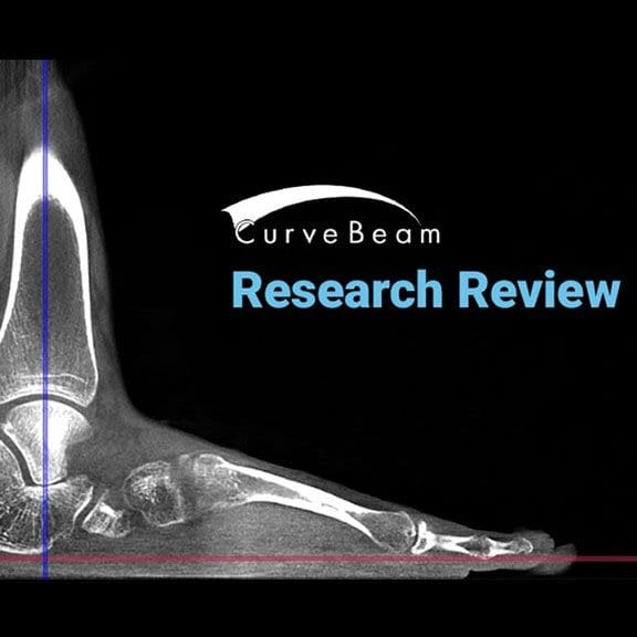
Orthopedic decision-making depends on accurate representation of anatomy—particularly when joint alignment and bone relationships change…

CT Has Numerous Advantages over X-Ray in the Taylor Spatial Frame Treatment
Winner of the Medical Device Excellence Award, the Taylor Spatial Frame (TSF), is regularly employed to treat complex fractures, bone deformities, and nonunion (http://www.smith-nephew.com/). Once TSF is implemented in a patient, a surgeon inputs information about the initial deformity into computer software that analyzes it and creates a “virtual hinge” of the deformity, the first and most important step in the creation of a corrective daily plan.
The TSF’s six-axis movement allows for precise corrections, but treatment can be imperfect without the correct input parameters in deformity, frame, mounting etc. CT is just as accurate as x-ray when scanning these parameters, but it is far more efficient—reducing errors and delays in treatment.
Following the initial correction, up to one-third of patients will experience persisting deformities known as residual deformities. These deformities can result from incomplete or insufficient corrections, which can be resolved by inputting new information about the deformity into the software that creates prescriptive correction plans. With that said, inaccuracies in the mounting parameters will nearly always result in unexpected translation-angulation deformities.
According to the study, “Calculating the Mounting Parameters for Taylor Spatial Frame Correction Using Computed Tomography,” by Kucukkaya et al. (2011), the exacting calculations of mounting parameters are integral to rapidly and effectively resolving residual deformities, and computed tomography (CT) techniques are uniquely precise in these calculations. Intraoperative fluoroscopy and postoperative x-ray techniques require that the effects of magnification be calculated.
If mounting parameters change during treatment, which they often do, recalculation is necessary. These issues make exact calculations nearly impossible. In contrast, Kucukkaya et al. (2011) found that CT calculations account for such changes and complexities, reducing residual deformities as well as treatment delay. CT’s advantages are particularly obvious in deformities with rotational elements, which are especially prone to deformities.
The Taylor Spatial Frame relies on the input of accurate data. The precision and flexibility of CT sets it apart as the best method for calculating the necessary parameters. The one drawback of CT is the relative increase in radiation exposure, though these effects are not uniform throughout the body and are negligible in knee and ankle cases. This minimal risk can be reduced even further by utilizing Cone Beam CT instead of the conventional narrow beam.
By using a wide beam in a cone shape, CurveBeam systems reduce the number of scan rotations required around the region of interest and thus the exposure to radiation. Similarly, the systems dramatically lower mA settings and the need for close positioning to the region undergoing imaging. This nearly eliminates CT’s radiation, its sole drawback in TSF. The Taylor Spatial Frame, when paired with CurveBeam cone beams, increases the effectiveness of deformity treatment while reducing the occurrence and persistence of residual deformities. This is accomplished with risk of radiation nearly eliminated.
To learn more about the advantages of cone beam CT in Taylor Spatial Frame treatment just visit Curvebeam online today!


