It is feasible for a single-practitioner podiatry practice to add weight bearing CT (WBCT) imaging and realize economical…
WBCT Indications Series: Midfoot Arthritis
Midfoot Arthritis
Midfoot arthritis occurs when there is loss of cartilage at the tarsometatarsal and/or navicular-cuneiform joints. It has the potential to cause significant pain, disability, and decreased quality of life. The most common etiology is posttraumatic degeneration, but primary degeneration and inflammatory diseases can also result in osteoarthritis of the midfoot joints.
A weight bearing CT scan can:
- Provide a clearer assessment of degenerative changes and joint space narrowing within the midfoot joints, which is normally impaired due to the natural overlap of adjacent midfoot bones viewed two-dimensionally with conventional radiography1.
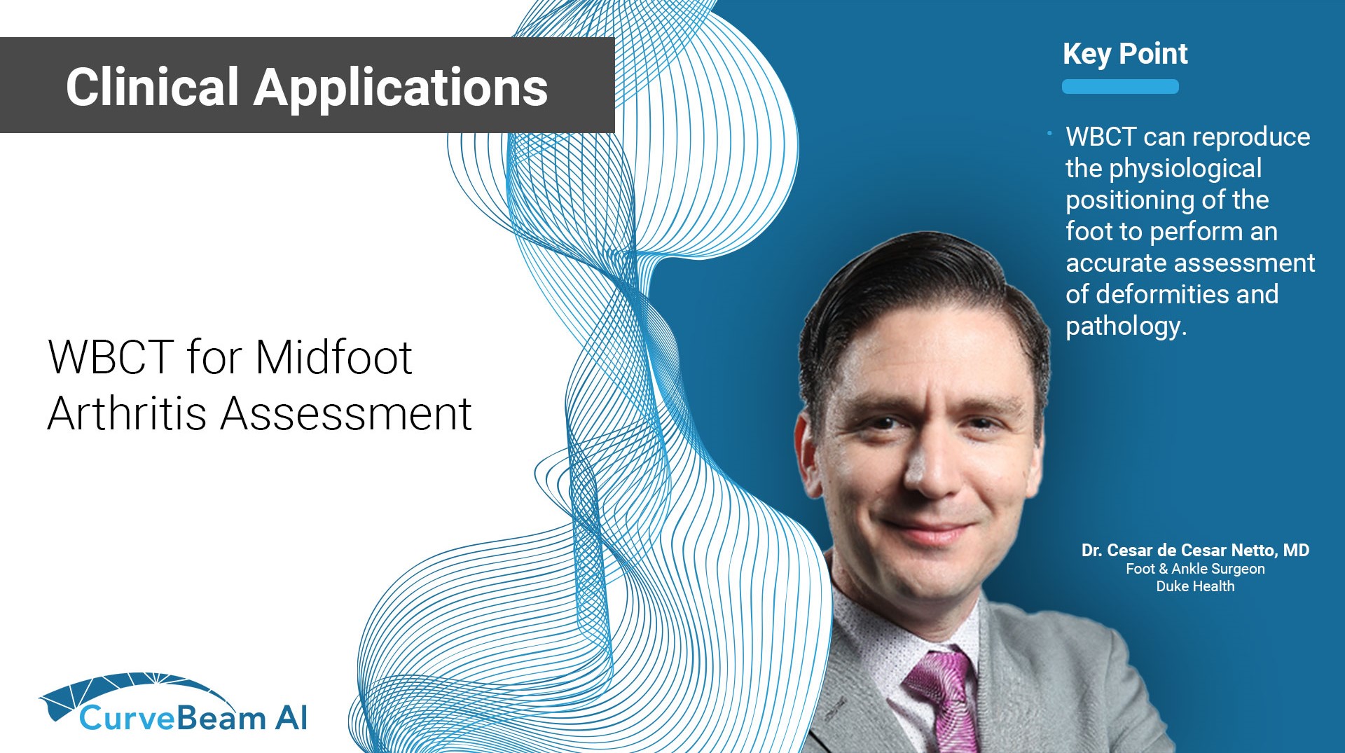
- Reproduce the physiological positioning of the foot to perform an accurate assessment of deformities and pathology2.
An accurate diagnosis and grading of the severity of osteoarthritic joints in the midfoot have been shown to be clinically relevant in treating the pathology early in its course and avoiding late-stage invasive procedures such as arthrodesis3.
In addition, WBCT better quantifies3 the structural deformity of Chopart, talonavicular, and calcaneocuboid joints when compared to conventional radiography and non-weight bearing computed tomography images.
Treatment Planning
Patients with unresolved symptoms after conservative treatment may be indicated for midfoot arthrodesis. A WBCT scan is an important tool in the planning and execution of midfoot surgical interventions. As a result of the complex structural anatomy, WBCT can help surgeons better visualize3 which joints may be pain generators and need to be included in the fusion.
In addition, the high reliability and reproducibility4,5 of WBCT images for the three-dimensional evaluation of the midfoot joints can help the foot and ankle surgeon determine the amount of correction required to appropriately realign the midfoot during surgery.
Postoperative Assessment
For postoperative evaluation of operative treatment of midfoot arthritis, a WBCT can:
- Accurately assess the adequacy of correction6.
- Accurately assess the healing process of midfoot joint fusions3.
- Act as a reliable method of identifying potential early failures in surgical treatment, which can help surgeons intervene earlier or plan a reoperation to obtain a favorable outcome6.
Midfoot Arthritis
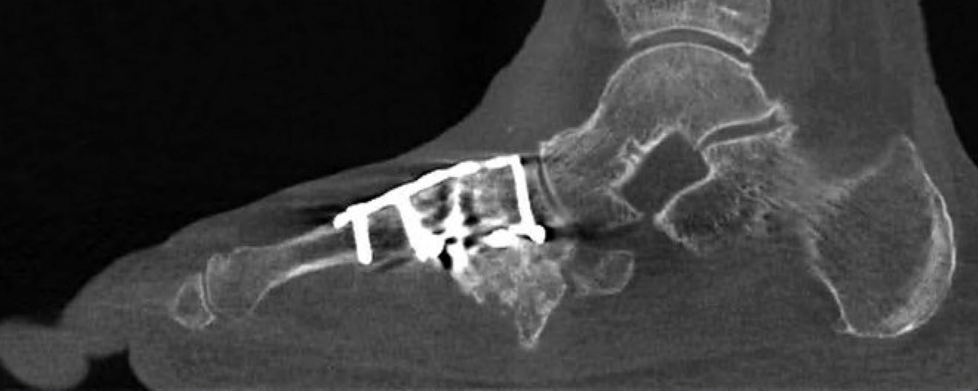
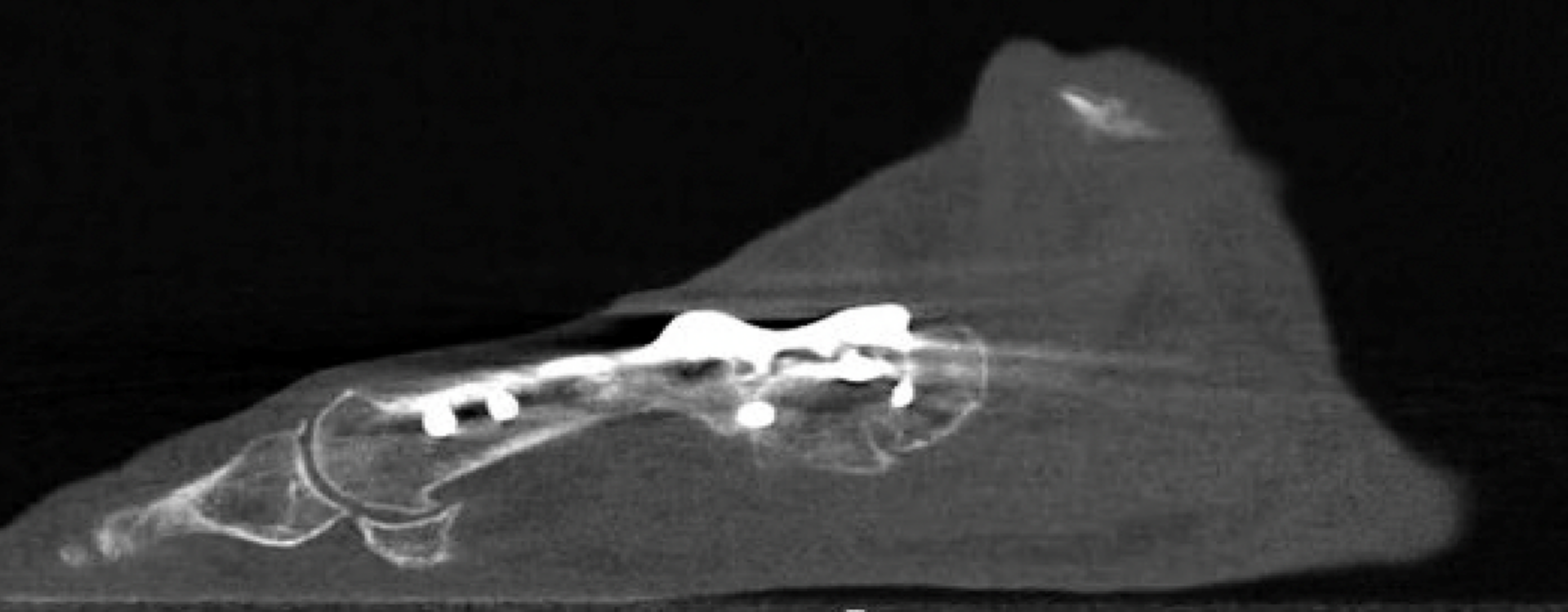
65 yo female with bilateral midfoot pain for 20 years, worse on the right side.
Images demonstrated severe degeneration of the midfoot joints. However, the severity of deformity and involved joints were difficult to determine on plain radiographs alone. As shown here, subluxation at the first and second TMT joints was better visualized on WBCT (bottom two scans) images than conventional radiographs (top scan).


Post-Op Assessment
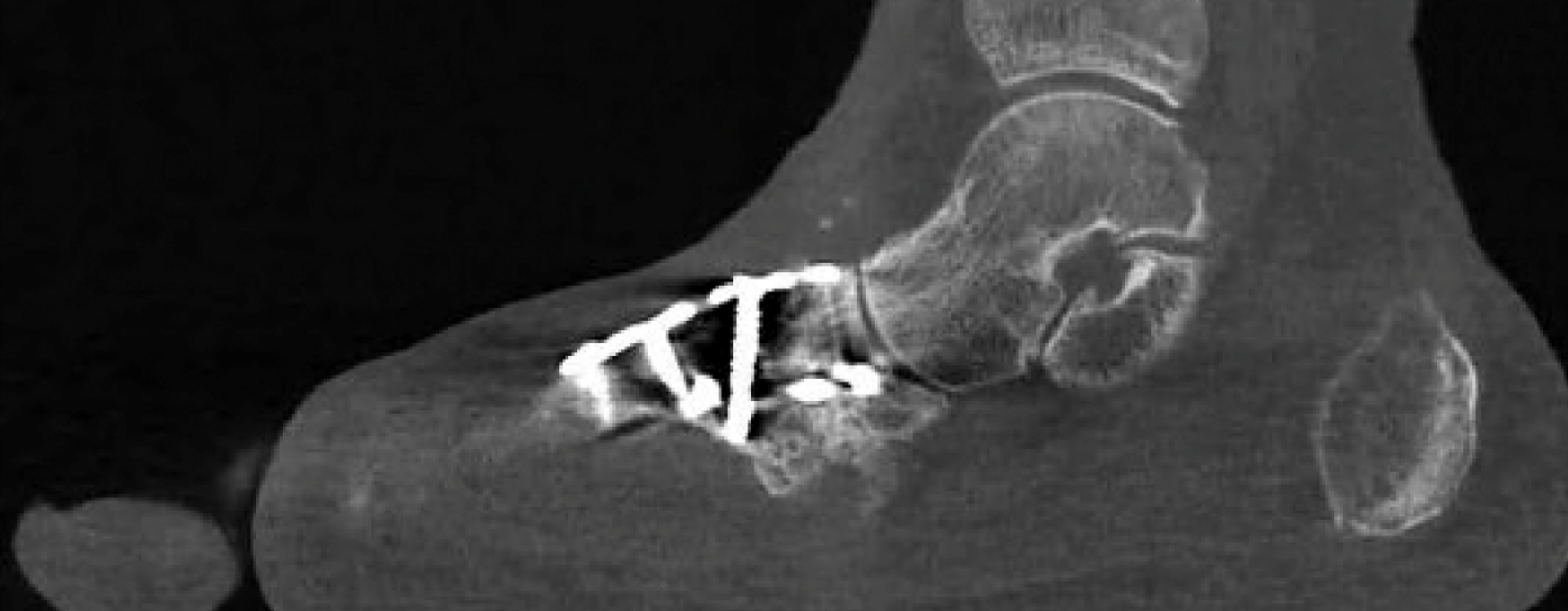
Obtaining a WBCT scan postoperative assists in the visualization of adequate restoration of the midfoot alignment, especially when surgical intervention was performed at multiple joints. Additionally, WBCT scans can assess bony bridging and fusion across the midfoot joints.
Click Here to get direct links to the latest studies using WBCT scans.

Dr. Cesar de Cesar Netto, MD
Dr. Cesar de Cesar Netto, a Duke Health Foot and Ankle Surgeon, has studied multiple pathologies of the foot and ankle, with focus on flatfoot deformity, Achilles tendinopathy and advanced imaging of the foot and ankle.
(1) de Cesar Netto C, Myerson MS, Day J, Ellis SJ, Hintermann B, Johnson JE, Sangeorzan BJ, Schon LC, Thordarson DB, Deland JT. Consensus for the Use of Weightbearing CT in the Assessment of Progressive Collapsing Foot Deformity. Foot Ankle Int. 2020 Oct;41(10):1277-1282. doi: 10.1177/1071100720950734. Epub 2020 Aug 27. PMID: 32851880.
(2) Jeng CL, Rutherford T, Hull MG, Cerrato RA, Campbell JT. Assessment of Bony Subfibular Impingement in Flatfoot Patients Using Weight-Bearing CT Scans. Foot Ankle Int. 2019 Feb;40(2):152-158. doi: 10.1177/1071100718804510. Epub 2018 Oct 8. PMID: 30293451.
(3) Dibbern KN, Li S, Vivtcharenko V, Auch E, Lintz F, Ellis SJ, Femino JE, de Cesar Netto C. Three-Dimensional Distance and Coverage Maps in the Assessment of Peritalar Subluxation in Progressive Collapsing Foot Deformity. Foot Ankle Int. 2021 Jun;42(6):757-767. doi: 10.1177/1071100720983227. Epub 2021 Jan 27. PMID: 33504217.
(4) Rojas EO, Barbachan Mansur NS, Dibbern K, Lalevee M, Auch E, Schmidt E, Vivtcharenko V, Li S, Phisitkul P, Femino J, de Cesar Netto C. Weightbearing Computed Tomography for Assessment of Foot and Ankle Deformities: The Iowa Experience. Iowa Orthop J. 2021;41(1):111-119. PMID: 34552412; PMCID: PMC8259196.
(5) Ortolani, M., Leardini, A., Pavani, C. et al. Angular and linear measurements of adult flexible flatfoot via weight-bearing CT scans and 3D bone reconstruction tools. Sci Rep 11, 16139 (2021). https://doi.org/10.1038/s41598-021-95708-x
(6) Steadman J, Sripanich Y, Rungprai C, Mills MK, Saltzman CL, Barg A. Comparative assessment of midfoot osteoarthritis diagnostic sensitivity using weightbearing computed tomography vs weightbearing plain radiography. Eur J Radiol. 2021 Jan;134:109419. doi: 10.1016/j.ejrad.2020.109419. Epub 2020 Nov 21. PMID: 33259992.

