It is feasible for a single-practitioner podiatry practice to add weight bearing CT (WBCT) imaging and realize economical…

WBCT Indication Series: End Stage Ankle Arthritis
End Stage Ankle Arthritis
End-stage ankle arthritis occurs when there is loss of cartilage at the tibiotalar joint. Post-traumatic ankle arthritis is the most common etiology.
A weight bearing CT scan can:
- Allow for accurate assessment of malalignment and congruency of the tibiotalar joint1.
- Evaluate the position and degenerative changes at adjacent joints including the subtalar and talonavicular joints1.
- Assist in planning deformity correction of the foot and hindfoot (e.g. COFAS type 3 and 4 cases)2.
- Provide a better understanding of the location of cysts and quality of the remaining bone3.
- Be used with the Stryker Prophecy® Surgical Planning preoperative navigation tool.
A WBCT scan can provide information about the severity of tibiotalar joint arthritis and cystic changes within the hindfoot. Importantly, a WBCT provides a three-dimensional assessment of any foot and ankle deformities for surgical planning purposes.
For patients with a painful total ankle replacement, a WBCT scan can be the first and only test to evaluate for possible causes of pain such as gutter impingement or malalignment of the ankle replacement or foot4.
Treatment Planning
A WBCT scan can be used to plan for concomitant correction of foot deformities (e.g. a heel slide, stabilization of the medial column). It can also help to identify degenerative changes in adjacent joints, which may need to be addressed at the same time or in a staged fashion. WBCT scans can also assess congruency of the tibiotalar joint.
For TARs requiring revisions, WBCT can help plan for realignment of the foot while sparing the implant and/or recognize the need for debridement of the gutters.
Postoperative Assessment
For postoperative assessment of operative treatment of ankle arthritis, a WBCT can:
Accurately assess sites of impingement and pain5.
Follow the size and locations of periprosthetic cysts.
Predict the likelihood of periprosthetic cyst formation based on varus/valgus3.
Surgical Planning for TAR
65 yo female with a history of a left talus fracture who developed significant hindfoot pain and deformity. Radiographs demonstrated tibiotalar arthritis as well as adjacent-joint arthritis.
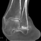
The calcaneal position was difficult to determine on X-Ray.
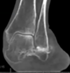
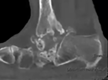
A WBCT scan showed that the talus was fractured and the calcaneus was subluxated laterally.
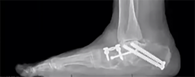
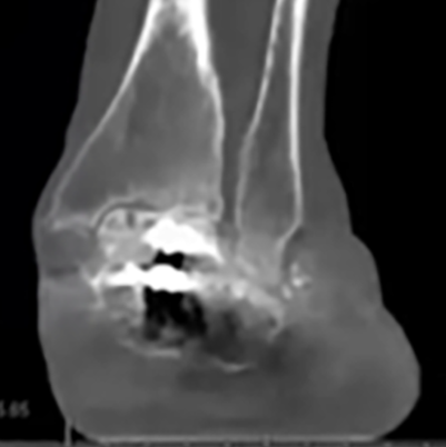
The patient underwent a staged triple arthrodesis. A post-operative WBCT showed insufficient reduction, which the treating surgeon planned to address in the second stage at time of total ankle replacement.
Postoperative Assessment
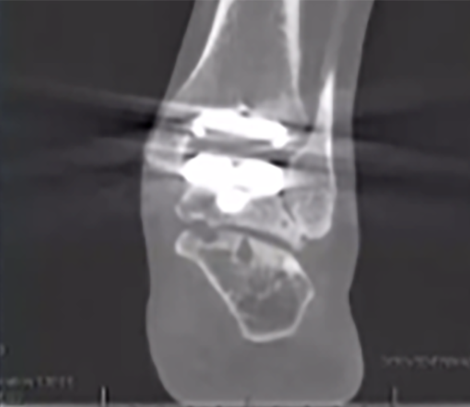
52 yo with post-traumatic degenerative joint disease of the ankle.
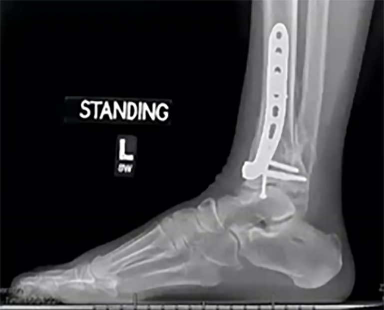
NWB CT after staged hardware removal did not reveal source of ankle pain.
WBCT after total ankle replacement showed clear lateral impingement, distally and at the level of the talar component.
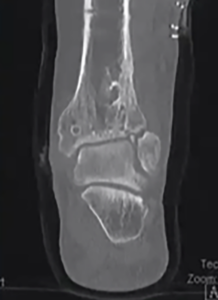
Treating surgeon performed a lateral gutter decompression and patient reported that pain went away.
Click Here to get direct links to the latest studies using WBCT scans.

Dr. Hodges Davis, MD
Dr. Hodges Davis, an OrthoCarolina Foot and Ankle Surgeon, has over 37 years of medical experience. His clinical interests include ankle arthritis, neuropathic disease, forefoot reconstruction, and deformity.
(1) Richter M, de Cesar Netto C, Lintz F, Barg A, Burssens A, Ellis S. The Assessment of Ankle Osteoarthritis with Weight-Bearing Computed Tomography. Foot Ankle Clin. 2022 Mar;27(1):13-36. doi: 10.1016/j.fcl.2021.11.001. Epub 2022 Jan 31. PMID: 35219362.
(2) Kvarda P, Heisler L, Krähenbühl N, Steiner CS, Ruiz R, Susdorf R, Sripanich Y, Barg A, Hintermann B. 3D Assessment in Posttraumatic Ankle Osteoarthritis. Foot Ankle Int. 2021 Feb;42(2):200214. doi: 10.1177/1071100720961315. Epub 2020 Oct 17. PMID: 33073607.
(3) Lintz F, Mast J, Bernasconi A, Mehdi N, de Cesar Netto C, Fernando C; International Weight-Bearing CT Society, Buedts K. 3D, Weightbearing Topographical Study of Periprosthetic Cysts and Alignment in Total Ankle Replacement. Foot Ankle Int. 2020 Jan;41(1):1-9. doi: 10.1177/1071100719891411. Epub 2019 Nov 28. PMID: 31779466.
(4) Kim JB, Park CH, Ahn JY, Kim J, Lee WC. Characteristics of medial gutter arthritis on weightbearing CT and plain radiograph. Skeletal Radiol. 2021 Aug;50(8):1575-1583. doi: 10.1007/s00256-020-03688-2. Epub 2021 Jan 7. PMID: 33410964.
(5) Jeng CL, Rutherford T, Hull MG, Cerrato RA, Campbell JT. Assessment of Bony Subfibular Impingement in Flatfoot Patients Using Weight-Bearing CT Scans. Foot Ankle Int. 2019 Feb;40(2):152-158. doi: 10.1177/1071100718804510. Epub 2018 Oct 8. PMID: 30293451.

