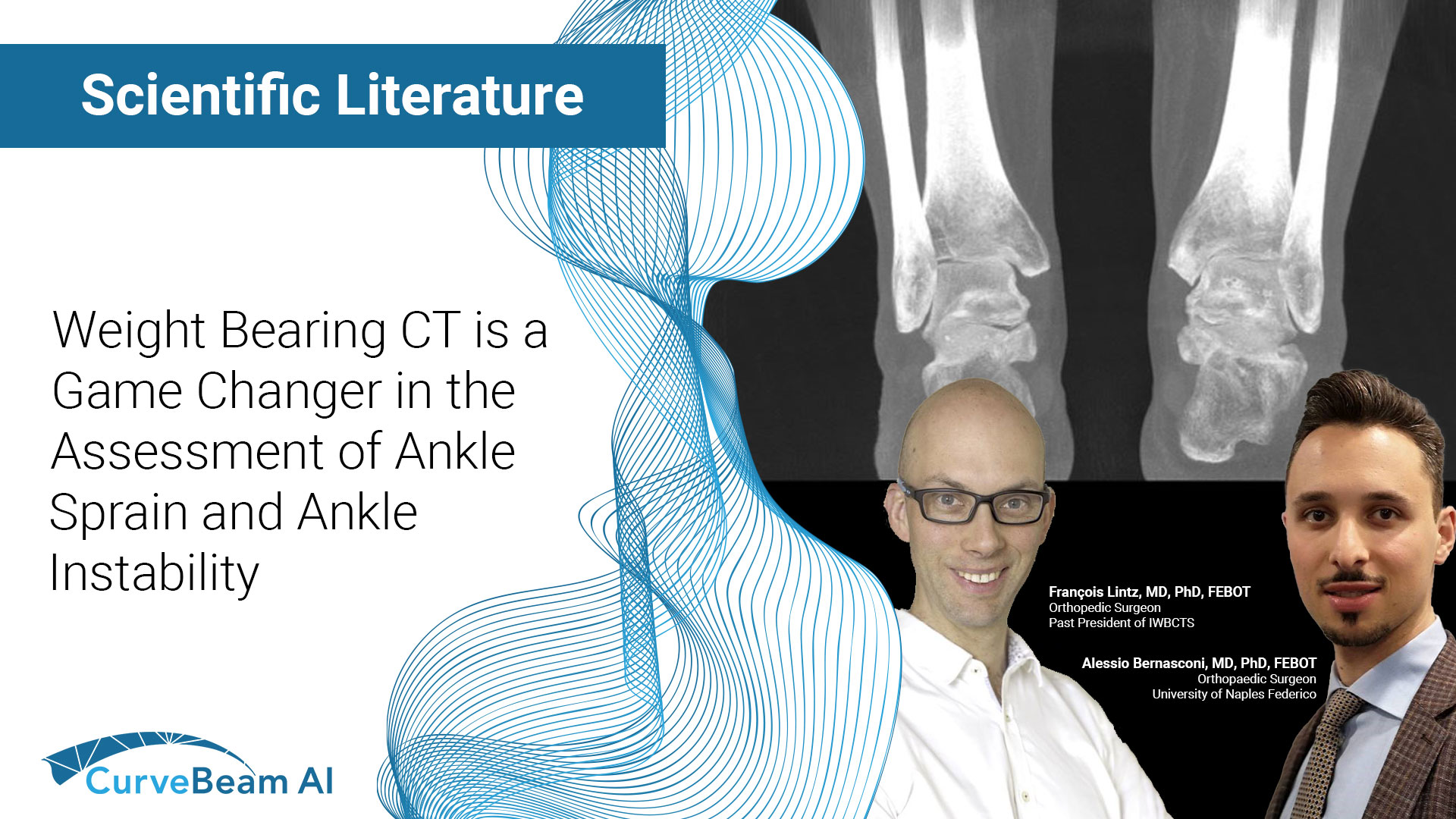
Orthopedic decision-making depends on accurate representation of anatomy—particularly when joint alignment and bone relationships change…

Can WBCT Be a Game-Changer in the Assessment of Ankle Sprain and Ankle Instability?
Key Points:
- The most important advantage of weight bearing CT (WBCT), which utilizes cone beam CT (CBCT) technology (a three-dimensional (3D) imaging modality) is immediate access to 3D images, resulting in faster and better diagnostic capabilities.
- Up to 40% of missed diagnoses of ankle trauma associated fractures could theoretically be avoided with primary use of WBCT in accident and emergency departments.
Diagnosing Ankle Sprains in the Emergency Department
Ankle sprain (AS), which represents roughly 20-30% of all lower limb trauma and 60-70% of all ankle trauma, is the most common reason for emergency department visits around the world. Since it is considered a relatively benign condition, clinicians do not often give enough attention to patients presenting with an AS injury, increasing the risk of misdiagnosis. Orthopedic cone beam CT (CBCT) has a huge potential to improve diagnosis and reduce waiting times, in turn freeing up conventional CT (MDCT) for other health issues.
Ankle sprains (AS) and chronic lateral ankle instability (CLAI) are complex conditions that are challenging to treat. CBCT is an innovative imaging modality with a body of supporting literature reporting reduced radiation exposure, shortened examination time and operating time, and a decreased time interval between injury and diagnosis.
Researchers analyzed the limited literature available on using CBCT as a primary modality for diagnosing AS and CLAI. They found:
- WBCT provides 3D imaging with radiation doses similar to conventional 2D radiographs (2DXR), though 2DXR often requires additional views to uncover hidden lesions caused by bone superimpositions.
- CBCT generally has better spatial resolution than MDCT , and is indicated for suspicion of bone lesions; including fractures, osteochondral lesions, small avulsion lesions, and subtle joint dislocations. Detecting such lesions, which often go undiagnosed after AS “is where CBCT could be “is most useful in the emergency department” if “systematically used as a primary means of investigation.”
A Deeper Look at Chronic Ankle Instability
When it comes to CLAI, literature is even more scarce, but researchers were able to glean these findings:
- Researchers have hypothesized for years that there may be a correlation between bone alignment, especially varus hindfoot, and the risk of developing CLAI . WBCT has proven reliable and accurate in measuring hindfoot alignment by enabling 3D measurements of the talar tilt and rotation, which has been found in the past to be a significant component of tibiotalar instability.
- WBCT’s ability to provide bilateral comparative imaging is informative when the comparative side can be considered devoid of pathology, or with at least clinical asymmetry.
- Advanced softwares can use WBCT data to calculate bone axes and map the distance between joints to objectively quantify talar tilt. These software tools could eventually help with initial clinical staging of the condition, providing pre- and post-surgical comparisons, and in patient follow-up evaluations.
Summary
Researchers summarized that there is an immense opportunity for future research on the impact of WBCT and CBCT in diagnosing, evaluating, and treating AS and CLAI. Early studies have shown the effect on management of foot and ankle trauma, particularly from a cost saving and clinical management perspective.
Researchers added that WBCT and CBCT have already changed the understanding of ankle pathology and is about to revolutionize the management of ligamentous injuries and concomitant pathology.
To read the full study click here.


