Orthopedic decision-making depends on accurate representation of anatomy—particularly when joint alignment and bone relationships change…

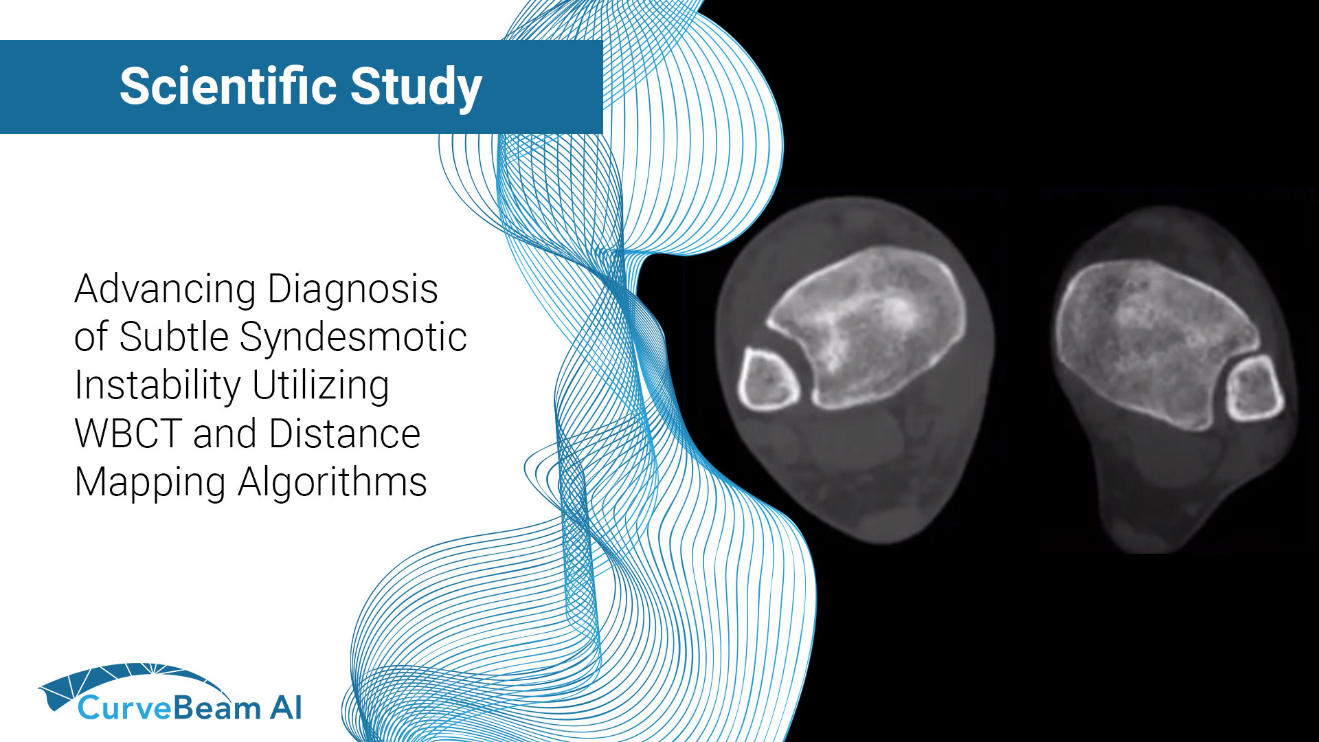
Cadaveric Diagnostic Study of Subtle Syndesmotic Instability Using a 3-Dimensional Weight Bearing CT Distance Mapping Algorithm
Key Points:
- Imaging modalities such as plain radiographs (X-Ray), computed tomography (CT), and magnetic resonance imaging (MRI), don’t have the diagnostic accuracy needed to detect syndesmotic widening or subtle instability.
- Weight Bearing CT (WBCT) imaging when combined with a distance mapping algorithm allows for a more accurate view of subtle syndesmotic instability.
The diagnosis of syndesmotic instability has been historically challenging, and chronically unstable injuries can potentially lead to ankle arthritic degeneration. Distal tibiofibular syndesmotic injuries occur in about 17% of ankle sprains and 20% of ankle fractures.
The diagnostic accuracy of different imaging modalities, such as X-Ray, CT, and MRI, in detecting syndesmotic widening still needs to be improved. WBCT has emerged as a promising diagnostic alternative as it could support the detection of even more subtle instabilities.
Up to now, no cadaveric study has been able to accurately diagnose subtle syndesmotic instability without the use of external rotational stress when there is no deltoid ligament injury. Dr. Cesar de Cesar Netto, MD, PhD, et al, out of the Department of Orthopedics and Rehabilitation, University of Iowa, Iowa City, Iowa, USA, set out to use a recently proposed 3D WBCT distance mapping algorithm to successfully diagnose this subtle instability.
Method
Nineteen matched pairs of through-the-knee cadaveric specimens (38 legs) were used in this study. Specimens were mounted in a frame that allowed simulated axial weight bearing. They were then scanned using CurveBeam AI’s HiRise WBCT system in the normal pre-injury state and after complete syndesmotic ligament sectioning. The deltoid was kept intact, and no external rotational stress was applied. Syndesmotic incisura and lateral gutter distances were assessed and compared between pre-injury ipsilateral, contralateral, and injured states using a 3D WBCT distance mapping algorithm. The receiver operating characteristic (ROC) curve and area under the curve (AUC) were calculated for the comparison of syndesmotic distance measurements between injured specimens and controls.
Results
Researchers found significantly increased distances in the injured specimens when compared with controls as well as average relative syndesmotic widening in injured specimens at the first 1, 3, 5, and 10cm (proximal to the apex of the distal tibial articular surface), respectively. Widening was more pronounced in the anterior aspect of the syndesmosis, where the diagnostic accuracy was found to be the highest at the first 1 and 3 cm of the syndesmotic incisura, with AUC values ranging from 80.9% to 83.0% and threshold diagnostic values of relative syndesmotic widening as low as 0.43 mm.
Conclusion
Researchers were able to successfully apply a 3D WBCT distance mapping algorithm to accurately detect subtle syndesmotic instability in specimens with complete syndesmotic ligament sectioning and an intact deltoid ligament, and in the absence of external rotational stress.
While there were limitations due to the cadaveric rigidity, cleaning sectioning of syndesmotic ligaments, and no traumatic energy involved, (more pronounced instability are expected in a clinical scenario) researchers hoped that the results of this study could stimulate and support the use of 3D WBCT distance mapping algorithms in resolving the complex riddle of accurately diagnosing true, subtle syndesmotic instability. It was noted that the application of this promising technique in a prospective and diagnostic cohort of patients with syndesmotic injuries and potential syndesmotic instability is paramount to confirming the importance of their findings.
To read the full study click here.


