AI-Driven Segmentation on Weight Bearing CT Introduction Accurate segmentation is critical for orthopedic workflows, including…
Avoiding False Negatives by Dodging Potential Pitfalls
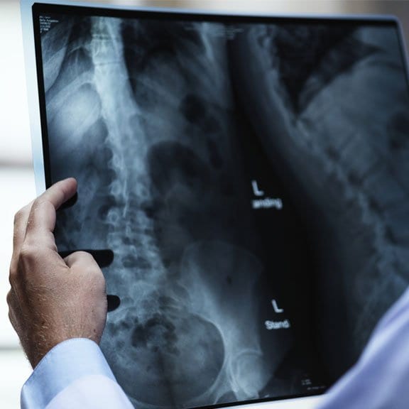
It would seem radiography reigns supreme when it comes to initial fracture detection. And yet, some researchers from the University of Washington’s Department of Radiology believe that relying on a single x-ray image can lead to a false negative. In their article Radiographic Pitfalls in Lower Extremity Trauma, authors Alice Ha, Jack Porrino, and Felix Chew examine the possible reasons for a missed diagnosis and measures that can be taken to avoid these inaccuracies. While the article lists a variety of pitfalls that can trip up radiologists, these can generally be broken down into three groups.
The first group relates to technological hitches. Detecting a fracture relies on having a variety of views, proper positioning, and technically sound equipment. While severe fractures may be identified from various viewpoints, others may only be detected when viewed from a specific angle, and fractures that go unnoticed can lead to more severe issues down the road. Additionally, the introduction of digital radiography has led to a belief that insufficient tube current can display an underexposed radiograph. To avoid false negative conclusions, there must be a thorough set of technical checks in place.
The second group that Ha, Porrino, and Chew focus on arise from physiological complications. Nondisplaced fractures, for instance, are often impossible to spot using a basic radiograph as the lack of displacement makes the bone appear intact, especially if weight bearing scans are not a part of the process. Utilizing a CT scan or an MRI can help avoid these errors, particularly in the lower extremities where there’s a higher possibility of missed fractures. And, according to the authors, fractures in places where hardware or artificial replacements have been installed can be all but invisible to radiographs, and thereby require a more comprehensive scan.
The third group of pitfalls highlighted in the article are attributed to human error. This can include everything from eliminating that one vital viewing angle during the imaging process, to simple faulty reasoning. A radiologist may identify a fracture, for example, but fail to realize the fracture is atypical for its location, and mis- or under- diagnose treatment for a more far-reaching issue. There is also the fact that many normal anatomic variations, such as sesamoid bones with multiple parts, can be mistaken for fractures. Other options must be implemented to account for human error.
At CurveBeam, we strive to erase potential pitfalls. Whether it’s allowing doctors to examine fractures through a weight bearing CT scan using our pedCAT technology, or giving a comprehensive look at a patient’s lower extremities with CurveBeam’s forthcoming LineUP, our products reduce false negative occurrences to ensure patients are getting the care they need.
To learn more about how CurveBeam products help to avoid radiographic pitfalls, visit https://curvebeamai.com/ today!
Related Posts


