Advancements in CT imaging are transforming orthopedic surgery, reducing surprises and improving surgical outcomes. In…
Conventional Measurements of OLTS with CT Grossly Overestimate the Size of Lesions
When orthopedic surgeons attempt to evaluate and manage osteochondral lesions of the talus (OLTs) using conventional computed tomography (CT), they may overestimate their size. This, in turn, influences treatment strategies. Therefore, OLTs often warrant advanced imaging studies, especially in revision or cases with cystic defects. To determine the effectiveness of these techniques, a study, “Value of 3D Reconstructions of CT Scans for Calcaneal Fracture Assessment,” was performed by Foot & Ankle International (FAI).
Understanding the True Anatomy and Size of OLTs May Help Guide Treatment
OLTs are defined as localized irregularities in the articular chondral surface also involving the subchondral bone. The exact cause of these lesions is unclear, but they are likely the result of a rotational ankle injury, such as a ligamentous sprain or bony fracture. OLTs are a common issue in orthopedic medicine; however, treatment remains a clinical challenge. Most OLTs can initially be managed with nonoperative treatment. When this approach fails, surgery is often indicated. Surgical intervention depends on the chronicity of the lesion, size of the lesion, presence of cystic lesions, and the stability of the cartilage. Many procedures for larger lesions require autograft or allograft tissue to fill the void, but these procedures can be very costly and do not always address the problem effectively. Understanding the true anatomy and size of these lesions may help better guide treatment, allowing for accurate preoperative planning and potential cost savings.
Conventional CT Scans Overestimate OLT Size
The results of the FAI study showed that conventional measurements of OLTs with CT scans grossly overestimated the size of the lesion. The average approximate OLT volumes from 10 independent observer CT measurements comprised of a rectangular prism (Vrec), a right cylinder (Vcyl), and right cone (Vcone) were compared to morphometric true-volume (MTV) via 3D reconstruction. On average, the Vrec, Vcyl, and Vcone estimates were 303%, 864%, and 285% greater, respectively, than the MTV. The FAI study demonstrates that volume measurements of OLTs using conventional CT typically overestimate true-volume approximations when compared to OLTs created with 3D reconstructions.
CurveBeam Imaging Provides Valuable Insights to Orthopedic Surgeons
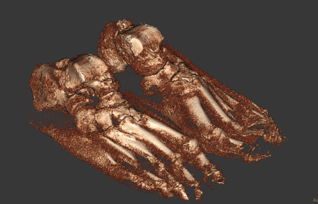
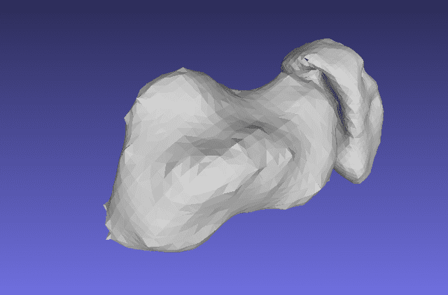
3D reconstructions of CT scans are used frequently in orthopedic surgery; however, the usefulness of this technique in OLTs has not been reported in the literature. 3D CT scans, such as those created using CurveBeam Cone Beam CT imaging technology, provide insight previously unavailable through either 2D and plain radiography. These detailed images, like those seen below, provide more precise measurements that can assist surgeons in both accurate diagnosing and preoperative planning.
Bilateral – Talar Head Exposed Sharp Filter: CubeVue, CurveBeam’s custom visualization software, can segment a volume to reveal joint surfaces, such as the talar head.
Talus Navicular Cluster: CurveBeam’s proprietary segmentation tools (under development) are demonstrated here using third-party viewing software.
CurveBeam designs and manufactures Cone Beam CT imaging equipment for the orthopedic and podiatric specialties. CurveBeam’s pedCAT system and CubeVue custom visualization software have been installed in facilities across the United States, Europe, Australia, and China. More recently, CurveBeam’s InReach system, a multi-extremity CT optimized for hand, wrist, and elbow imaging, and LineUP system, a weight-bearing multi-extremity CT for imaging of the foot, ankle, and knee, have been added to the product line. Today, CurveBeam Cone Beam CT scans are setting new standards in orthopedic 3D imaging worldwide.
To find out more, visit https://www.curvebeamai.com/about/about-curvebeam/.
[1] https://www.researchgate.net/publication/324808166_The_Role_of_3D_Reconstruction_True-Volume_Analysis_in_Osteochondral_Lesions_of_the_Talus_A_Case_Series

