Chat with us, powered by LiveChat Skip to content

Jon-David Deeson Testimonial on pedCAT

During OrthoForum’s Annual Conference in early March, OrthoTennessee CEO Jon-David Deeson discussed his experiences with the pedCAT, our low-dose, in-office bilateral weight- bearing foot and ankle CT scanner. His clinic in eastern Tennessee has been using the pedCAT for just…

Read more

Weight Bearing CT Imaging for Cuboid Subluxations

Dr. Michael Chin, DPM, presented how weight bearing CT imaging has changed how he evaluates cuboid subluxations at the American Academy of Podiatric Sports Medicine meeting held near the West Point Military Academy campus in early September, 2015. Dr. Chin began using the pedCAT in his office in February of this year. Not much research is out there on how to use plain radiographs to measure cuboid subluxations, Dr. Chin said in his lecture, titled, “Cuboid Syndrome…The Other Side of Heel Pain.” Dr. Chin has tested using a bilateral oblique projection to understand the cuboid/ metatarsal relationship, and has been able to observe a slide between the head of the fourth metatarsal and the head of the cuboid. An MRI could be ordered to see the condition of the peroneal tendon, but the study would be limited because the scan would not be weight bearing, he said.. A traditional CT scan would provide great visualization of the bone, but would provide no information on anatomic alignment. The pedCAT weight bearing CT imaging system is excellent for evaluating stress fractures, sesamoids, periosteal changes, or anything medullar, Dr. Chin said. Another benefit is he can measure the exact degree of subluxation between the cuboid and the fourth metatarsal head. Dr. Chin displayed pedCAT images depicting pre and post-reduction views of a cuboid subluxation. pedCAT scan of a pre-surgical patient with cuboid subluxation. Dr. Chin was able to reduce the subluxation to 2.18 mm. Dr. Chin practices at The Running Institute in Chicago.
Read more

pedCAT: A Positive User Experience

“After using the pedCAT for a year, I am very impressed with how much I need it, and how many different things I use it for,” Dr. Bob Baravarian said at the 2015 Western Foot and Ankle Conference held in Anaheim, Cali. The California Podiatric Medical Association hosted the meeting at the end of June. Dr. Baravarian cited Hallux Rigidus as one example where pedCAT imaging has been helpful because he is able to properly assess the sesamoids. Dr. Baravarian confirmed his practice is realizing positive revenue streams as a result of the device.
Read more
ALT TEXT

Weight Bearing CT in Everyday Practice

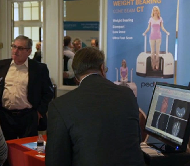
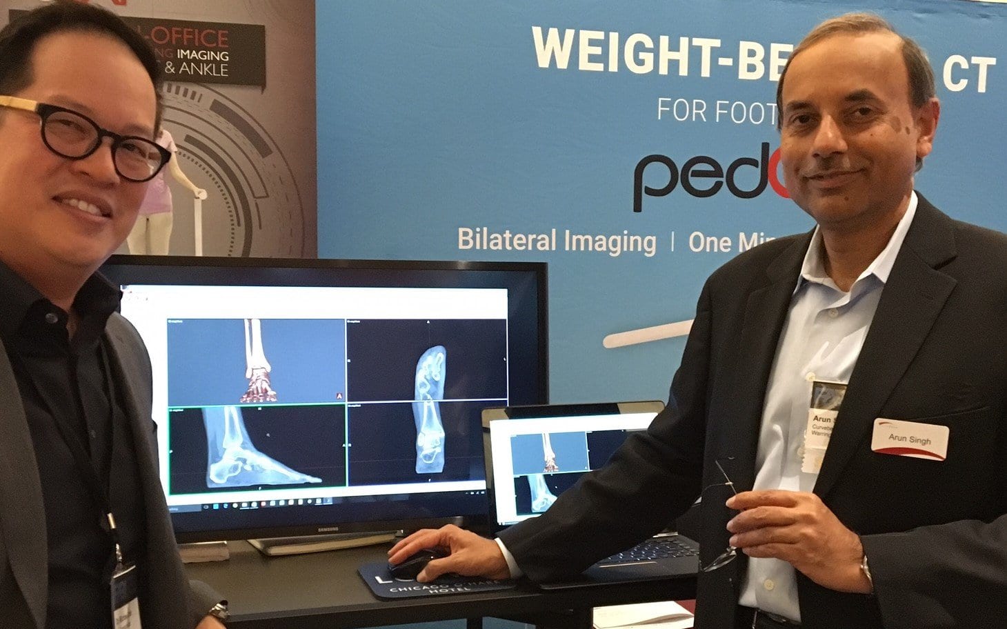
“In my opinion, this will be the standard of care in the next couple of years. We are switching from two-dimensional technology to three-dimensional technology and this is really the future of not just foot and ankle, but the future of medicine,” Dr. Alex Tievsky, DPM, said in a lecture he gave at the Graham International Implant Institute 8th Annual Symposium in Miami, on April 17. 2D is beginning to be phased out,” Tievsky said. “Now we’re beginning to see the problem from all angles and all planes, so this is super helpful from that respect.” 
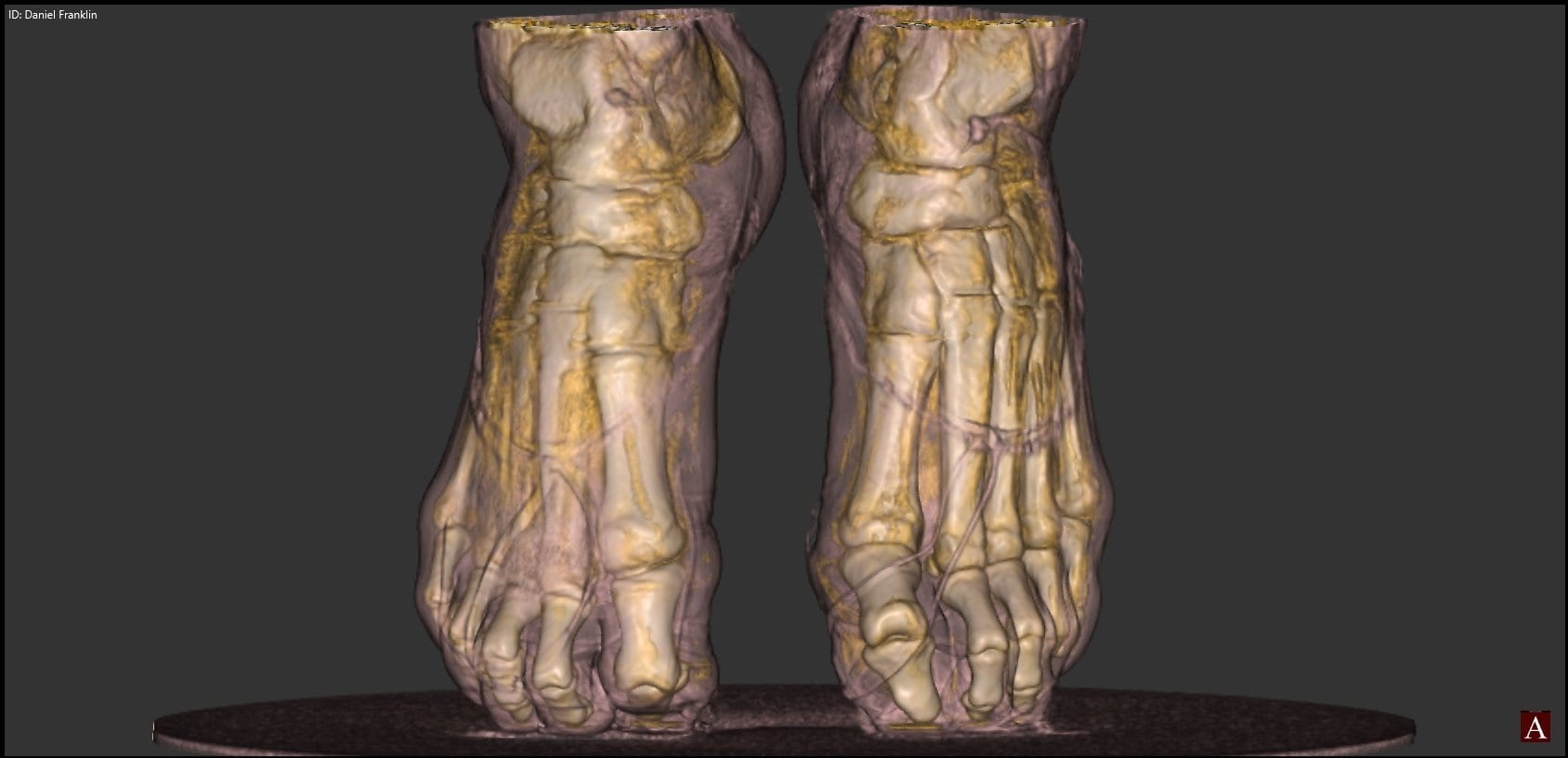
Dr. Tievsky presented a number of cases where he benefited from access to weight bearing 3D technology in his office. Clinical Case #1 
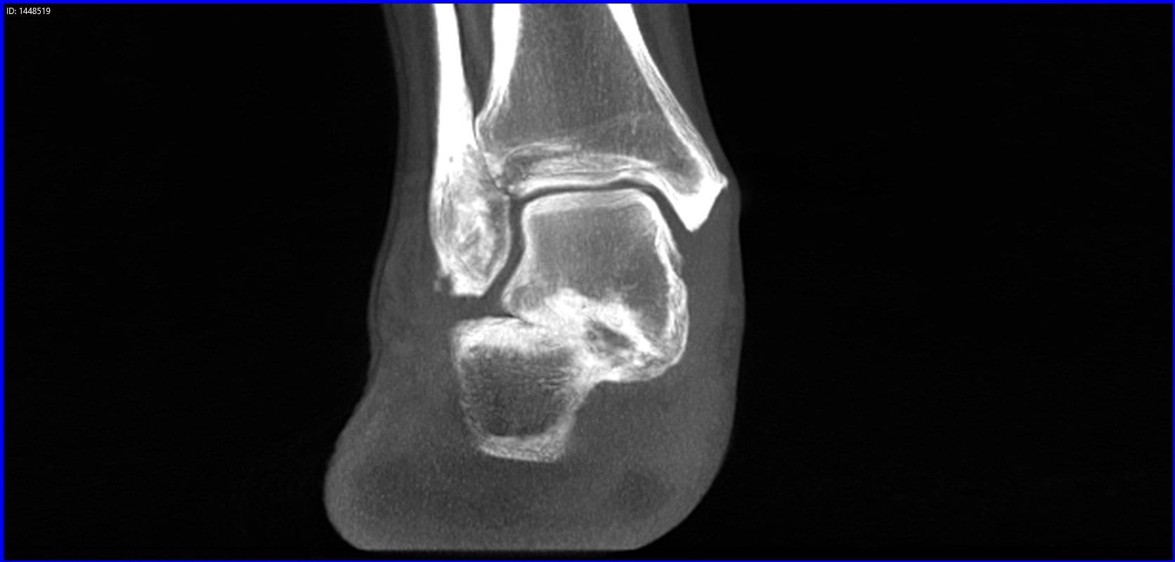
A 50-year-old female presented with bilateral flat feet for 20 years. She had heard about the HyProCure procedure, which corrects hindfoot misalignment through a minimally invasive procedure. She was eager to have the procedure done, no matter the cost. Dr. Tievsky took a pedCAT scan, and found she had a severe talar coalition. talar calc coalition coronal left “How many times do you catch a coalition on an X-Ray? It’s hard,” Dr. Tievsky said. “Sometimes you can see a halo sign, but it’s often missed. On the first visit, I was able to tell her, we either have to resect this coalition or we have to do a fusion. It’s impossible to get this level of information on an X-Ray.” Clinical Case #2 A 16-year old girl came in with first metatarsal head pain. She had already been to two other podiatrists in the last six months, and they had prescribed steroid injections. The pedCAT revealed a fracture on her fibular sesamoid that is extremely easy to miss on X-Ray. fractured sesamoid “Within her first 10 minutes in the office, we had a diagnosis,” Dr. Tievsky said. “We treated her appropriately. We immobilized her for eight weeks and gave her a bone stimulator. And she was pain free, three podiatrists later. She was happy, her mom was happy, and she never came back.” Clinical Case #3 A patient presented with a lateral plantar fasciitis, a talo-tarsal dislocation, back pain for five years, knee pain, and hip pain. A pedCAT revealed she had a tumor in her bone and it was eroding it. “There was no way we would have caught it on X-Ray,” Dr. Tievsky said. “We sent her out for oncology. It could have a malignant tumor, and we could have saved her life.” Cyst Coronal The scan is quick to take and you have a full work-up in about three minutes, Dr. Tievsky said as a closing statement. “This is a super important tool,” he said. “I’m kind of biased. I love this now. I can’t practice without it.”
Read more

Dr. Gary Briskin: The pedCAT is the Future

"It's the future - embrace it. It's your future - control it." Dr. Gary Briskin delivered this motivating message at the Foot & Ankle Business Innovations conference in Chicago on Jan. 23. Dr. Briskin, of University Foot & Ankle Institute in Santa Monica, Cali, discussed how the pedCAT weight bearing CT system has improved his practice since his group acquired one last year. His main take-away points were: - The pedCAT gives you the advantage when it comes to diagnosing pathology. "You can solve that Lisfranc case no one else can," Dr. Briskin said. "It opens up the midfoot, which historically has been a problem for us." - The pedCAT allows him to clearly determine if bones and joints are completely fused after surgery. "I do a lot of lapidus procedures," Dr. Briskin said. "My concern; is this patient fused adequately to start bearing weight?" The exact level of fusion is visible with a pedCAT scan 5 - 6 weeks later, and with minimal scatter. - Radiologists like the pedCAT images "I think I'm getting a better CT, because it's weight bearing and I think the quality is far superior. We get all of our CTs read by a radiologist. We have everything sent digitally. And the feedback we get is they are also impressed with the quality of the images."
Read more
Back To Top