Orthopedic decision-making depends on accurate representation of anatomy—particularly when joint alignment and bone relationships change…
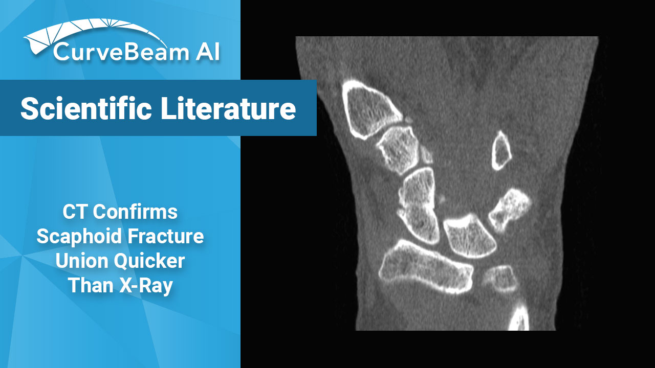
Cone Beam CT Confirms Scaphoid Fracture Union Quicker than X-Ray
Cone Beam CT (CBCT) is more accurate than X-Ray for predicting scaphiod healing at early follow-up (six weeks), thus enabling faster mobilization of patients and return to daily activity and work, according to a study published by Lucia Calisto et al.
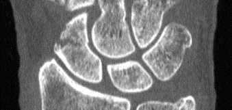
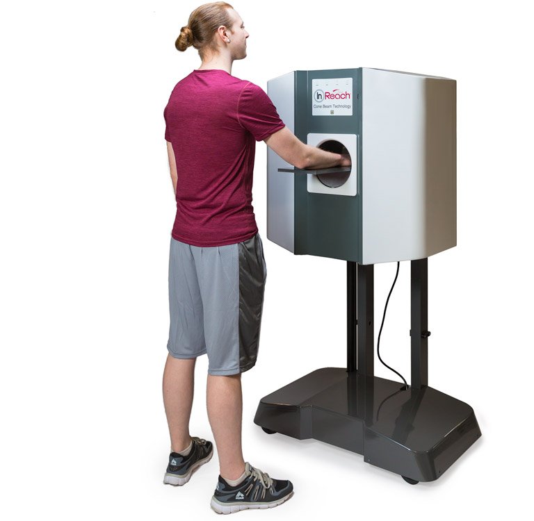
The CurveBeam AI InReach provides 0.2mm high resolution slices of the distal limbs.
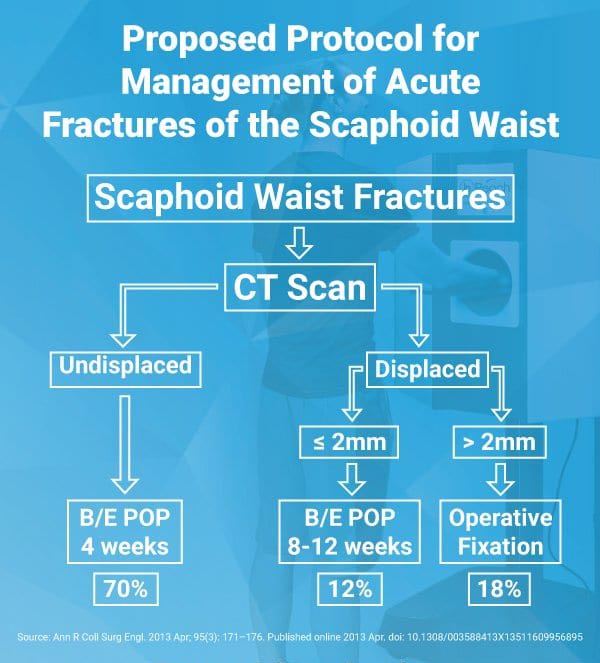
In a previous study, Professor Timothy Davis wrote CT studies demonstrate healing of a non-displaced fracture treated with a plaster cast can occur in as little as 4 weeks. If a fracture is displaced less than 2 mm, Davis said those CT studies suggest a plaster cast for 8 – 12 weeks.
CT is ideally performed for all scaphoid wrist fractures in the first week after injury to classify whether they are displaced or non-displaced, said Professor Davis, an orthopedic surgeon at Woodthorpe Hospital in Nottingham, UK said in his research paper.
Conventional medical CT is often ordered to confirm scaphoid healing, but involves significantly more radiation exposure to the patient than CBCT. CBCT’s lower dose could mean doctors could order repeat tests to monitor scaphoid healing, the study authers said.
By using CT as a baseline, researchers at the Roth/McFarlane Hand and Upper Limb Center in London said they were able to identify fractures which may have appeared non-displaced on X-Ray, but were actually minimally displaced.
“We feel that the added visualization of CT over plain radiography enables the surgeon to properly select which fractures are appropriate for non-operative cast treatment with an expected high degree of union,” the researchers said in a study published in The Open Orthopaedics Journal.
Out of the research setting, routine CT scans of scaphoid fractures may not be practically feasible, Professor Davis wrote.
“I appreciate that [routine CT assessment of scaphoid fractures] is impossible in many centers at the present time but it should become increasingly possible in the future,” Professor Davis wrote in the medical journal “Annals of the Royal College of Surgeons of England” in 2013.
The InReach is a compact CT imaging system dedicated to the hand, wrist and elbow. The system received FDA and CE approval in 2017. Since then, it has been installed in leading orthopedic centers and hospitals in the United States. The InReach allows orthopedic practices to offer CT imaging at the point-of-care.
“InReach has been an excellent asset allowing in-office imaging and rapid CT evaluation of the hands with complex diagnostic dilemmas,” said Dr. Lloyd Champagne, an orthopedic surgeon at the Arizona Center for Hand to Shoulder Surgery in Phoenix.
“Although the moment of follow-up regarding scaphoid healing remains a controversial topic, CBCT could shorten this period enabling the diagnosis of consolidation with higher confidence than x-ray. CBCT technology provides more reproducible results than X-Ray regardless of the experience of the radiologist or hand surgeon.” as stated in the 2020 Farracho et al study.
Fifteen percent of acute fractures of the scaphoid waist fail to unite if treated non-operatively in plaster, resulting in a persistent loss of function, according to the 2013 article. Plain X-Rays do not clearly show fracture features such as displacement and communition. Previous inter-observer studies have shown radiographs of scaphoid fractures are neither sensitive nor specific.


