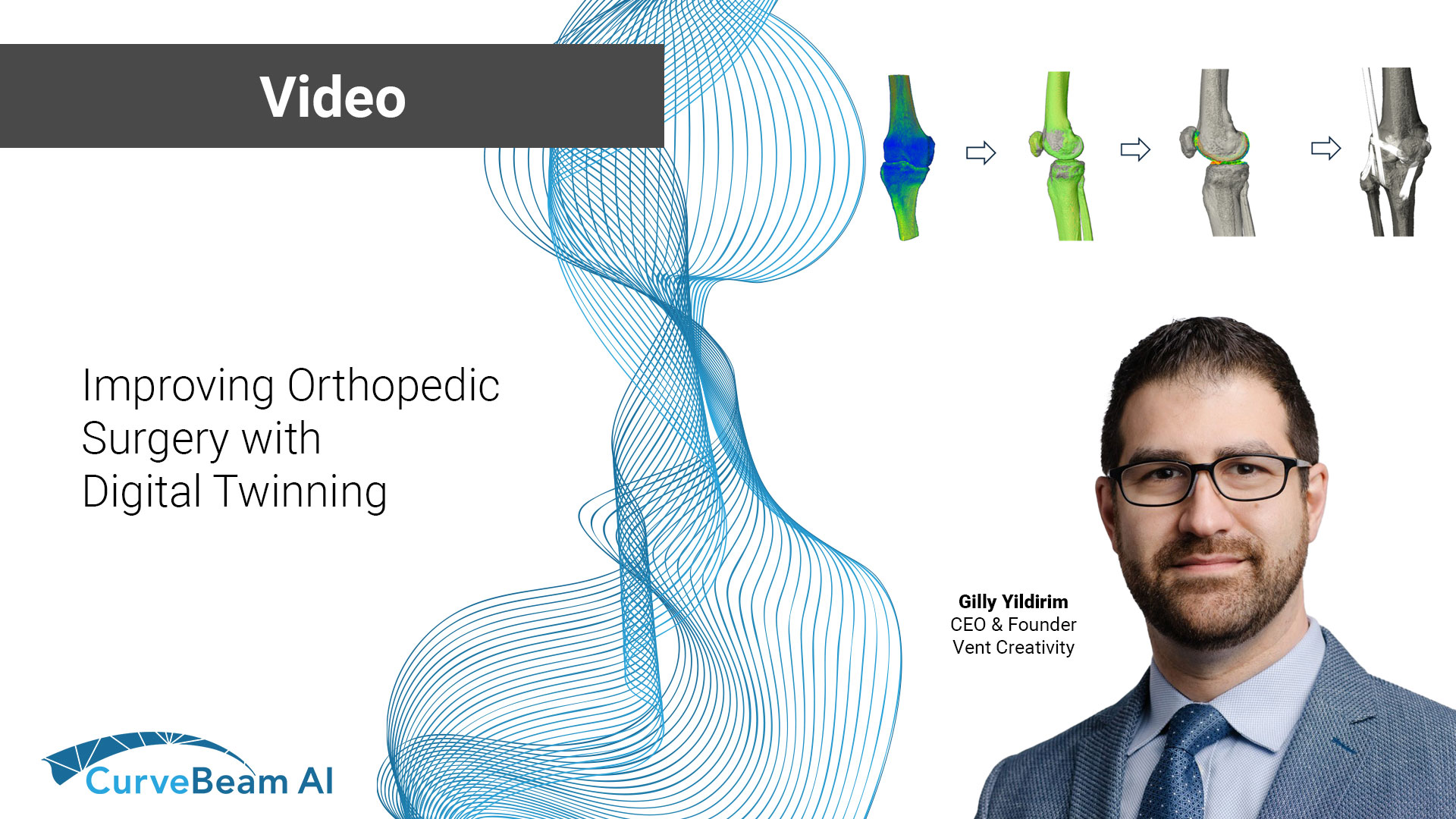
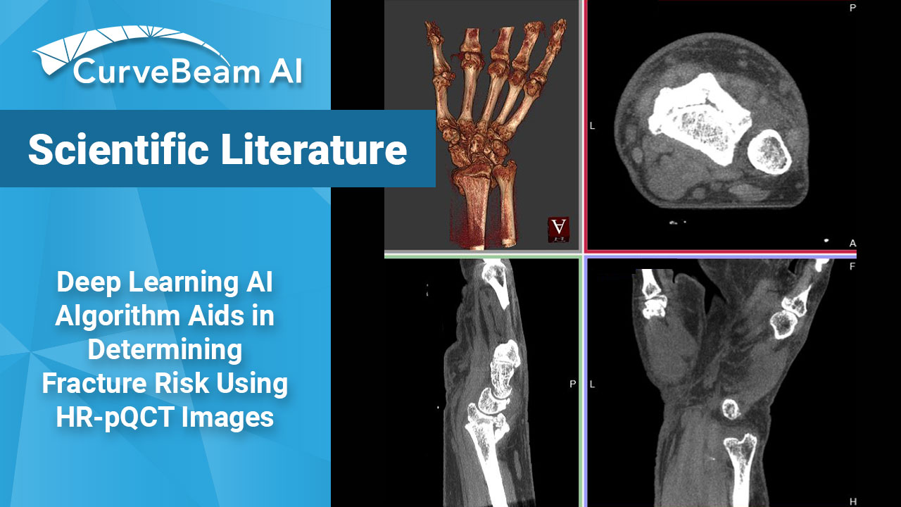
Advancements in CT imaging are transforming orthopedic surgery, reducing surprises and improving surgical outcomes. In…

Value of 3D Reconstructions of CT Scans for Calcaneal Fracture Assessment
Operative fracture treatment of irregularly shaped bones, such as the calcaneus, scapula and scaphoid, demands high-quality images of the area in question for both classification of the fracture and planning of the procedure. However, since plain radiographs do not provide enough pertinent information to this end, computerized tomography (CT) has become the standard for providing the necessary images for treatment of these irregularly shaped bones.
Yet, in a study observing 2D CT scans, only 42% of the evaluators were able to correctly classify the fracture, necessitating the need for some sort of change. Three dimensional CT renderings were proposed to fix the low inter-rater agreement generated by the 2D scans.
The foot in the video above was scanned on a pedCAT weight bearing CT imaging system for the foot and ankle. The calcaneus was segmented using CurveBeam’s CubeVue visualization software.
To evaluate the effectiveness of 3D CT scans, A standard set of CT secondary reformation scans were presented, followed by a questionnaire describing fracture anatomy and preoperative planning. Subsequently, 3D reconstructions were presented to the evaluates followed by the same questionnaire. After presentation of the 3D images, 49% of the evaluators changed their plan in regard to the approach and 29% in regard to the implants.
Five different data sets (four intra-articular and one extra-articular fractures) were presented to 57 evaluators. All groups, except that of surgeons with more than 20 years of experience, benefited from 3D CTs (Friedman test; P < .01). Inexperienced surgeons benefited more than experienced surgeons and complex fractures more than simple fractures. Specifically, regions of interest such as the middle facet and fractures extending into the calcaneo-cuboid joint were evaluated more precisely.
In regard to 3D CT scans, Böhmer1 described the topographic relationship between the fragments and the surrounding structures as useful for evaluating calcaneal fractures and for preoperative planning because the fractures can be seen from unusual perspectives. Likewise, Choplin2,3 posited 3D scans assist diagnosing foot deformities since the scans improve comprehension of the anatomy, particularly for especially complex fractures. For such complex fractures and anatomy, Pate5 evaluated 202 patients with complex musculoskeletal problems and found 3D CT particularly helpful. Others came to similar conclusions.
When comparing techniques for diagnosing foot deformities, Allon and Mears4 compared plain radiography, 2D CT, and 3D CT of 30 fractured calcanei and concluded that 3D CT improves preoperative planning and the choice of an adequate approach.
Overall, 3D CT scans provide insight previously unavailable through both 2D and plain radiography, which inexperienced surgeons tend to find more helpful in diagnosing and preoperative planning.
References
- Bohmer G, Roesgen M, Hierholzer G. Three-dimensional computerized tomography in trauma surgery. A case presentation [in German]. Aktuelle Traumatol. 1992;22(2):47-56.
- Choplin RH, Buckwalter KA, Rydberg J, Farber JM. CT with 3D rendering of the tendons of the foot and ankle: technique, normal anatomy, and disease. Radiographics. 2004;24(2):343-356.
- Choplin RH, Farber JM, Buckwalter KA, Swan S. Threedimensional volume rendering of the tendons of the ankle and foot. Semin Musculoskelet Radiol. 2004;8(2):175-183
- Allon SM, Mears DC. Three dimensional analysis of calcaneal fractures. Foot Ankle. 1991;11(5):254-263
- Pate D, Resnick D, Andre M, Sartoris DJ, et al. Perspective: three-dimensional imaging of the musculoskeletal system. AJR Am J Röntgenol. 1986;147(3):545-551.


