Orthopedic decision-making depends on accurate representation of anatomy—particularly when joint alignment and bone relationships change…
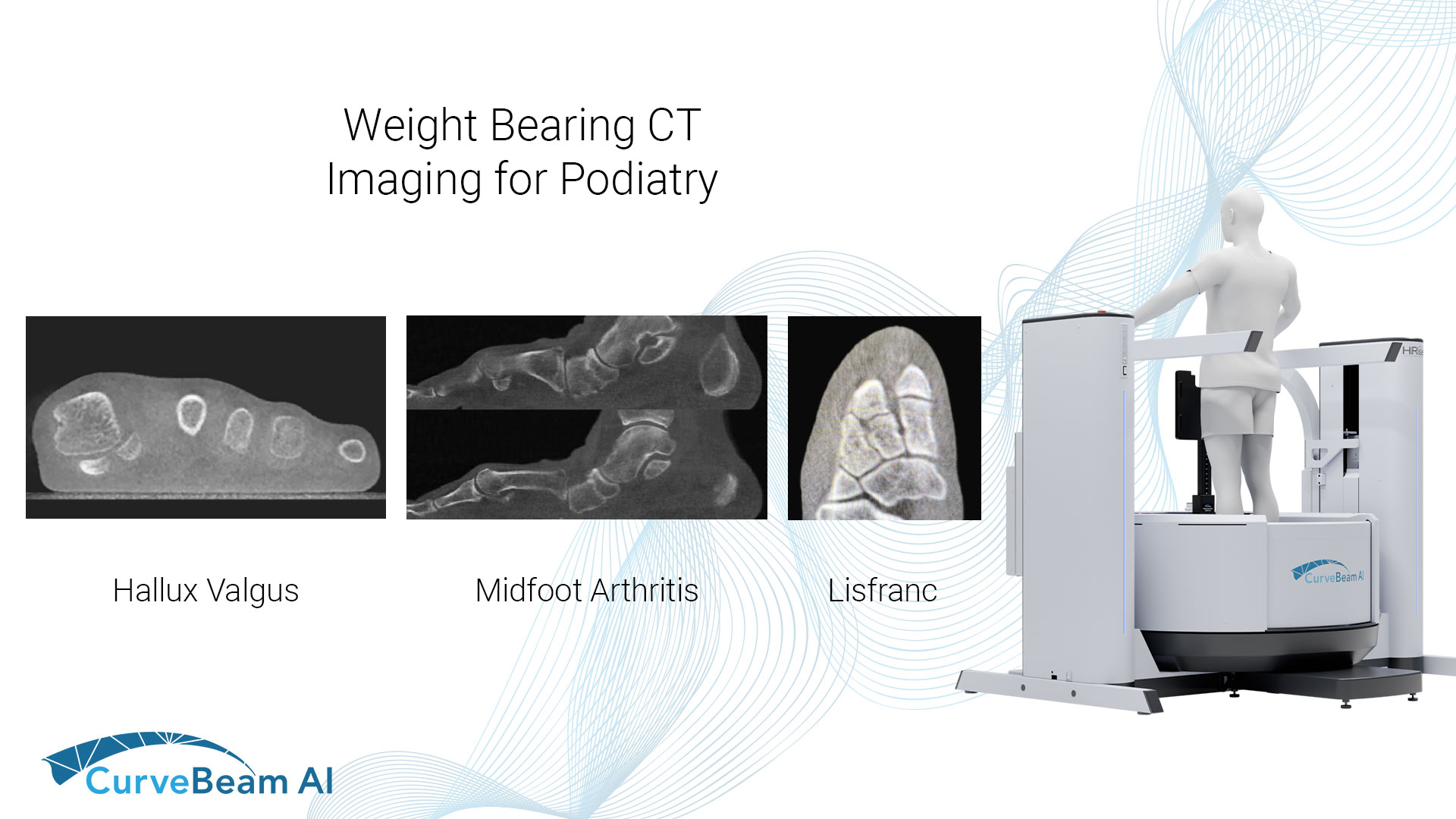
Weight Bearing CT Imaging for Podiatry
Common Indications
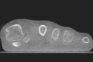
Hallux Valgus
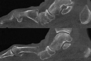
Midfoot Arthritis
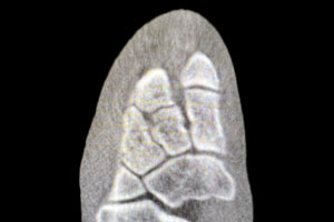
Lisfranc Injuries
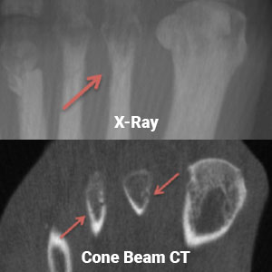
Fractures

“A plain X-Ray in many cases will not show small fractures in their early stages of presentation. For this reason, they are often misdiagnosed and mistreated. A low dose Cone Beam CT scan of the foot is very valuable in these situations.”
David J. Soomekh, DPM
Sports Medicine & Reconstructive Foot & Ankle Surgery
Foot & Ankle Specialty Group, Beverly Hills, CA
(1) Sripanich Y, Steadman J, Krähenbühl N, Rungprai C, Mills MK, Saltzman CL, Barg A. Asymmetric lambda sign of the second tarsometatarsal joint on axial weight-bearing cone-beam CT scans of the foot: preliminary investigation for diagnosis of subtle ligamentous Lisfranc injuries in a cadaveric model. Skeletal Radiol. 2020 Oct;49(10):1615-1621. doi: 10.1007/s00256-020-03445-5. Epub 2020 May 11. PMID: 32394072.
(2) Sripanich Y, Weinberg M, Krähenbühl N, Rungprai C, Saltzman CL, Barg A. Change in the First Cuneiform-Second Metatarsal Distance After Simulated Ligamentous Lisfranc Injury Evaluated by Weightbearing CT Scans. Foot Ankle Int. 2020 Nov;41(11):1432-1441. doi: 10.1177/1071100720938331. Epub 2020 Aug 20. PMID: 32819160.
(3) Lange, B., & Voldby, H. (2022, February 24). Webinar recap: WBCT scans of potentially unstable. CurveBeam AI. Retrieved March 30, 2023, from https://curvebeamai.com/webinars/webinar-recapwbct-scans-of-potentially-unstable-weberbser2-fractures/
Related Posts