When CT Is Not Enough In a recent discussion on the clinical value of weight…
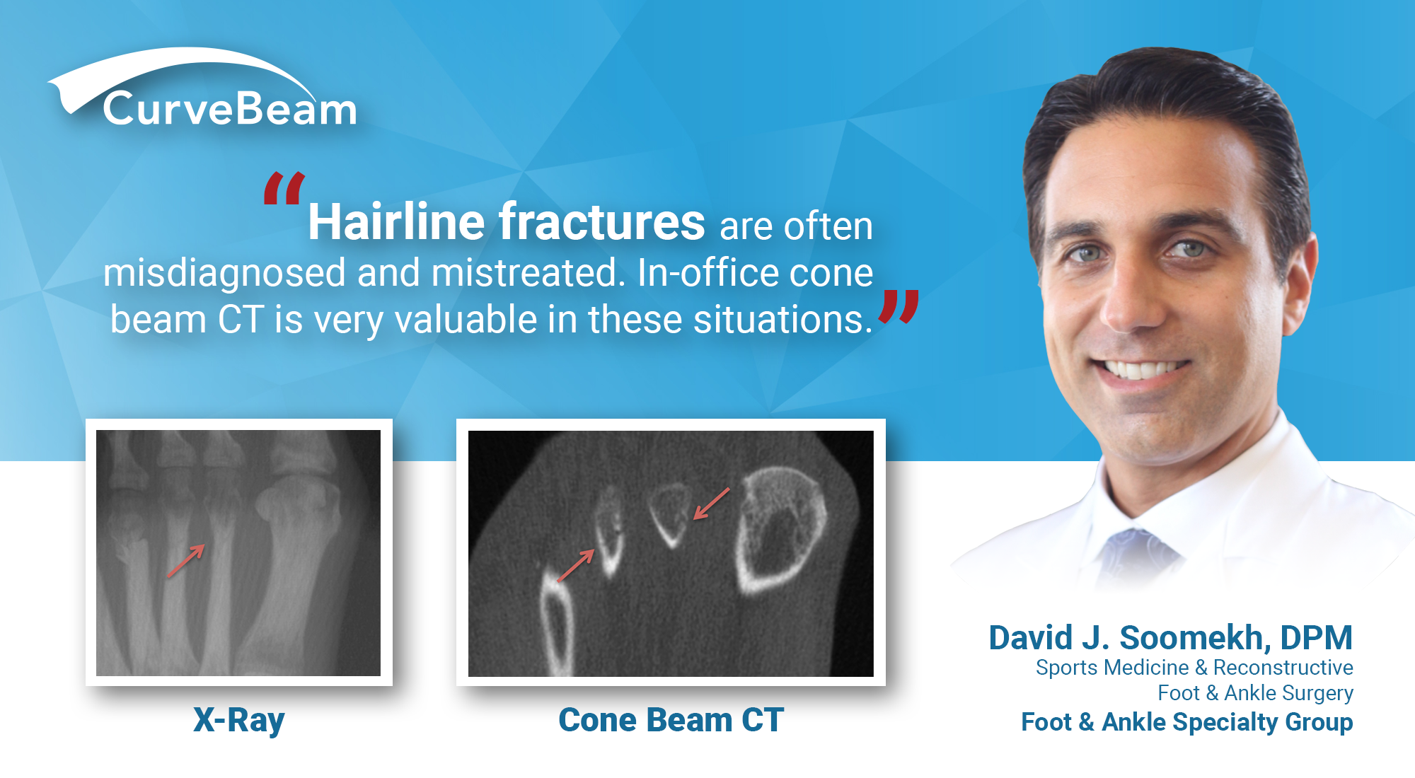
Hairline Fractures: Dx w/ In-Office CT Imaging

By Dr. David J. Soomekh, DPM, FACFAS
Guest Contributor
“I think I have a hairline fracture of my foot.”
“I hurt my foot, but it can’t be broken, because I can still walk on it.”
“I may have fractured my foot, but I hope it is not broken.”
“I just rolled my foot and I think I just sprained it.”
These are frequent comments we hear from patients when they present with an acute foot injury. There are common misconceptions about bone injuries in the foot and ankle. It is important to note that any disruption of the outer shell or the inner marrow of the bone is a broken bone. A “fracture” is a broken bone. A “hairline fracture” is a broken bone. A “stress fracture” is a broken bone. They are all simply different degrees of a break to the bone, and may warrant different treatments.
Stress fractures and hairline fractures of the foot or ankle are very common. They can occur when abnormal stress put upon the bone. This could be from increased walking or running, a new exercise routine, a rotational injury, or high impact fall. A recent example from current events is when President Elect Joe Biden slipped while playing with his dog.
Patients will often tell me they had a minor roll of the foot and or ankle when performing an activity or taking a misstep. When the foot rolls inward against the ground, there is undo pressure on the bone from the forces of the body over the foot and the forces of the ground pushing back against the foot. There is also the extra pull of the ligaments and tendons that attach to the bone that can injure the bone. These forces may stress the bone to its initial limitations causing a breach of the outer “shell” of the bone called the “cortex.”
It is a common misconception that one cannot walk on a broken bone. The patient will present with a dull and achy pain of the foot that they are able to walk on with a slight limp and pain. They will also notice some swelling that has been consistent since the injury. They may have been walking on the injured foot for many days hoping the pain would go away on its own before they present to the doctor. The physical examination would show tenderness on palpating the bone and perhaps movement of the bone.
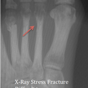
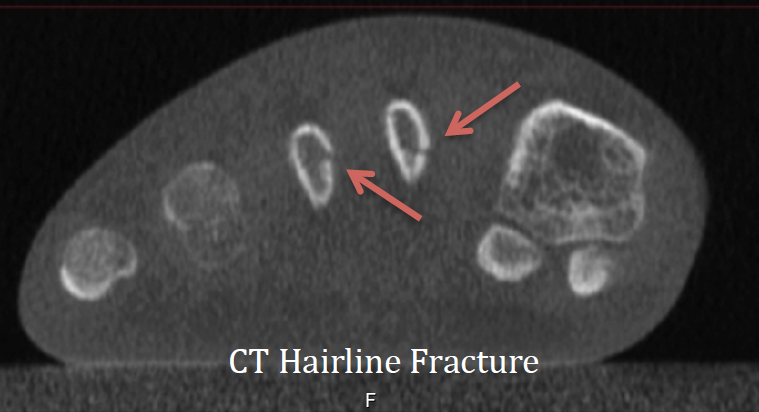
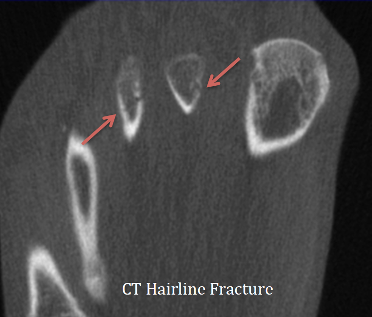
A plain X-ray of the bone in many cases will not be able to show these small fractures in their early stages of presentation. For this reason, they are often misdiagnosed and mistreated. An in-office low dose Cone Beam CT scan of the foot is very valuable in these situations. This type of imaging can scan the bones layer by layer in 3 dimensions with far superior accuracy and visualization than a plain X-Ray. Fractures not seen on plain X-Ray will be seen on this type of CT scan. Early diagnosis and treatment are the key to proper healing and an optimal return to activities and lifestyle.

President Elect Joe Biden’s initial X-Rays did not show any obvious fracture, his physician said in a statement. He was able to get a follow-up CT shortly after, which confirmed a hairline fracture.
Click here to listen to a CurveBeam Connect podcast episode on how weight bearing CT became Dr. Soomekh’s primary in-office modality.
Related Posts