Orthopedic decision-making depends on accurate representation of anatomy—particularly when joint alignment and bone relationships change…
Weight Bearing CT Imaging for Sports Medicine
Common Indications
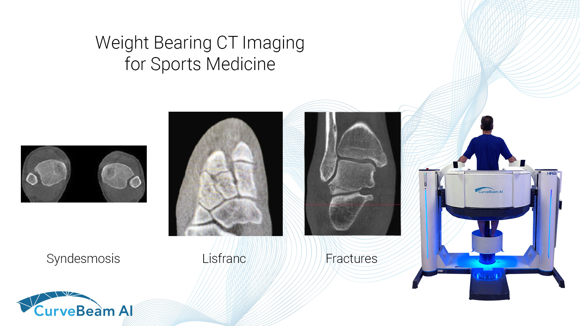
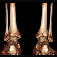
Syndesmosis
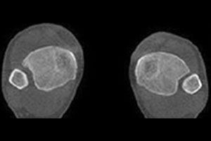
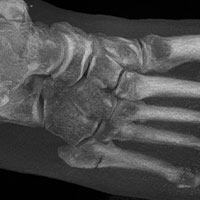
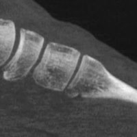
Lisfranc Injuries
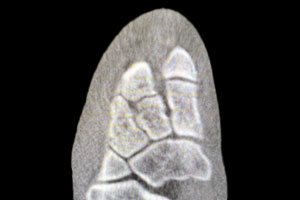
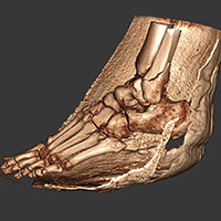
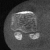
Fractures
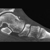

“I now CT every ankle fracture, and I have been surprised at the variability.”
Dr. Martin O’Malley, MD
Team Orthopedist
Brooklyn Nets, New York, NY
(1) Lintz F, Bernasconi A, Ferkel EI. Can Weight-Bearing Computed Tomography Be a Game-Changer in the Assessment of Ankle Sprain and Ankle Instability? Foot Ankle Clin. 2023 Jun;28(2):283-295. doi: 10.1016/j.fcl.2023.01.003. PMID: 37137623.
(2) Hagemeijer NC, Chang SH, Abdelaziz ME, Casey JC, Waryasz GR, Guss D, DiGiovanni CW. Range of Normal and Abnormal Syndesmotic Measurements Using Weightbearing CT. Foot Ankle Int. 2019 Dec;40(12):1430-1437. doi: 10.1177/1071100719866831. Epub 2019 Aug 23. PMID: 31442094
(3) Sripanich Y, Weinberg M, Krähenbühl N, Rungprai C, Saltzman CL, Barg A. Change in the First Cuneiform-Second Metatarsal Distance After Simulated Ligamentous Lisfranc Injury Evaluated by Weightbearing CT Scans. Foot Ankle Int. 2020 Nov;41(11):1432-1441. doi: 10.1177/1071100720938331. Epub 2020 Aug 20. PMID: 32819160.
(4) Bhimani R, Sornsakrin P, Ashkani-Esfahani S, Lubberts B, Guss D, De Cesar Netto C, Waryasz GR, Kerkhoffs GMMJ, DiGiovanni CW. Using area and volume measurement via weightbearing CT to detect Lisfranc instability. J Orthop Res. 2021 Nov;39(11):2497-2505. doi: 10.1002/jor.24970. Epub 2021 Jan 6. PMID: 33368556.
(5) Lange, B., & Voldby, H. (2022, February 24). Webinar recap: WBCT scans of potentially unstable. CurveBeam AI. Retrieved March 30, 2023, from https://curvebeamai.com/webinars/webinar-recapwbct-scans-of-potentially-unstable-weberbser2-fractures/
(6) Posadzy M, Desimpel J, Vanhoenacker F. Cone beam CT of the musculoskeletal system: clinical applications. Insights Imaging. 2018 Feb;9(1):35-45. doi: 10.1007/s13244-017-0582-1. Epub 2018 Jan 4. PMID: 29302798; PMCID: PMC5825310
Related Posts