Orthopedic decision-making depends on accurate representation of anatomy—particularly when joint alignment and bone relationships change…
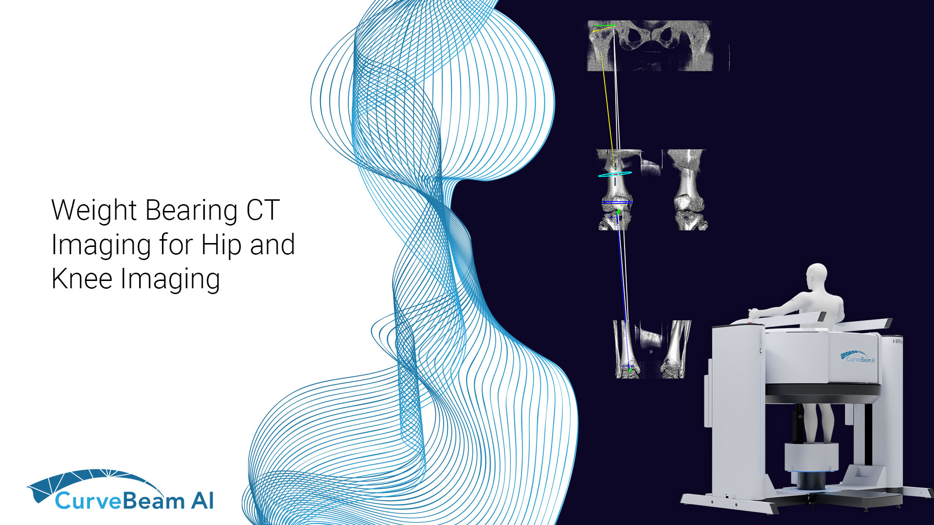
Weight Bearing CT for Hip & Knee Imaging
Common Indications
Osteotomy Planning
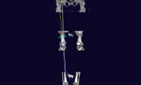
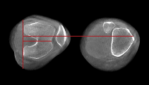
Patellar Instability
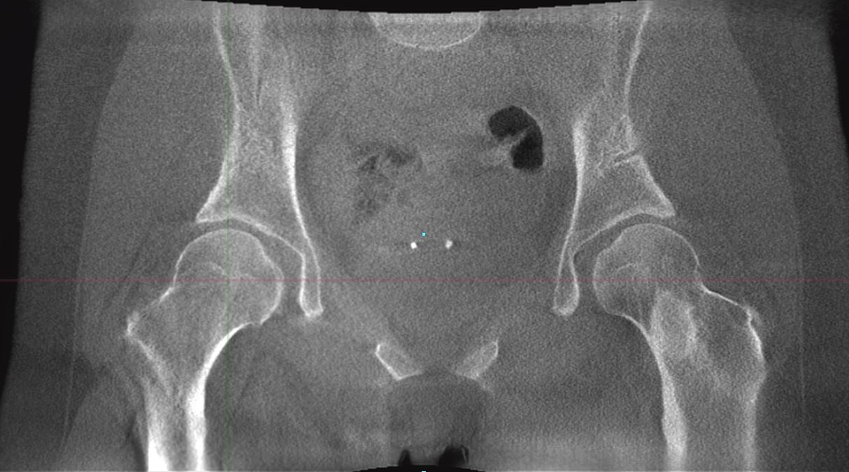
Hip Dysplasia
The Weight Bearing Difference
A 58-year-old patient with significant ankle pain sought a consultation with Dr. Walther. The patient had seen several orthopedic surgeons, one of whom had prescribed orthotics. However, the orthotics did not alleviate her pain. Her supine medical CT (MDCT) scan denoted only minimal arthritis. The patient could not get an MRI due to her pacemaker. Dr. Walther ordered a weight bearing CT (WBCT) scan, which revealed significantly reduced joint space. He performed a successful total ankle replacement on the patient three months later.
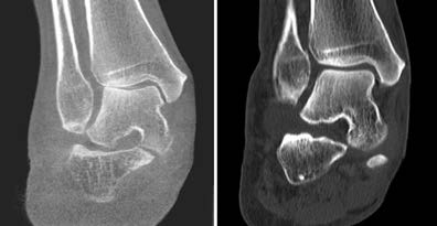
Cone Beam CT vs Supine CT

“A WBCT exam allows a completely new evaluation of many deformities. It allows axis misalignments, joint instabilities, or osteoarthritis to be better classified.”
Dr. Markus Walther, MD
Foot & Ankle MD
(1) Hirschmann, A., Buck, F.M., Fucentese, S.F. et al. Upright CT of the knee: the effect of weight-bearing on joint alignment. Eur Radiol 25, 3398–3404 (2015). https://doi.org/10.1007/s00330-015-3756-6
(2) Lullini G, Belvedere C, Busacca M, Moio A, Leardini A, Caravelli S, Maccaferri B, Durante S, Zaffagnini S, Marcheggiani Muccioli GM. Weight bearing versus conventional CT for the measurement of patellar alignment and stability in patients after surgical treatment for patellar recurrent dislocation. Radiol Med. 2021 Jun;126(6):869-877. doi: 10.1007/s11547-021-01339-7. Epub 2021 Mar 3. PMID: 33660189; PMCID: PMC8154791.
(3) Willey, M. & University of Iowa. (n.d.). Measuring Hip Instability in Dysplastic Patients with Weight Bearing CT. Orthopedic Research Society 2022 Annual Meeting, Tampa, Florida, United States of America.


