pedCAT: Improved Outcomes
The message at RSNA Annual Meeting this past November was clear: new US healthcare laws mean the practice of radiology is no longer about volume, but value.
“We are focusing on quality metrics. It’s becoming important for us to become champions of quality in our institutions,” said Dr. Vijay Rao, MD, in a course at the meeting.
How could a pedCAT add value to your practice?
We might get an idea by looking at a comparable new technology. Breast tomosynthesis mammography provides 3D imaging for breast cancer screening, similar to the way the pedCAT provides 3D imaging for the foot and ankle. Breast tomosynthesis technology can detect breast cancers earlier than traditional 2D mammography, and can more accurately pinpoint the size, shape and location of abnormalities, according to the Massachusetts General Hospital Imaging Department.
Dr. Liane Philpotts, professor of diagnostic radiology at the Yale School of Medicine, called tomosynthesis a “game changer” and a “win-win.”
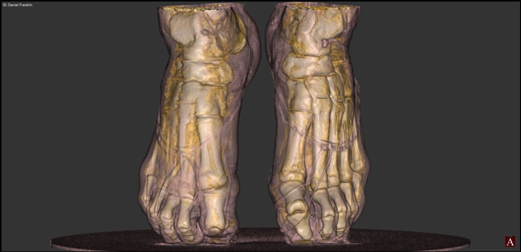
In the same way, the pedCAT eliminates variability, helps lead to better diagnoses, and makes both you and your patients more confident that treatment will result in better outcomes.
Dr. Erik Nilssen, MD, said the pedCAT helps him determine exactly when to allow patients to ambulate, “based on our ability to monitor fracture healing and fusion rates.”
Also, pedCAT scans can take the guesswork out of hindfoot alignment, said Dr. Martin O’Malley, MD, because they allow for reproducible measurements.
“We’ve never had a reproducible measurement,” until now, he said.
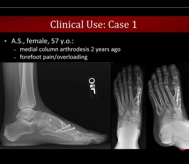
To see an example of a pedCAT scan that led to a more accurate diagnosis, click on the blog post title.