WBCT on the Program Thursday, January 22 11:10 am – 12:30 pm Symposium 2: Diagnostic…

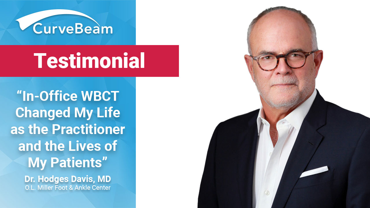
Testimonial: Dr. Hodges Davis, MD
Dr. Hodges Davis, MD said in the future, weight bearing CT will be the standard of care for all foot and ankle injuries and pathologies.
WBCT has become a routine part of his practice for fracture assessment and fusion evaluation, Dr. Davis said.In the video above, he describes how important it is to see the foot in 3D alignment.
“In-office WBCT changed my life as the practitioner and the lives of my patients,” he said. Here’s how:
- “I don’t make mistakes,” Dr. Davis said. If a fracture looks questionable on X-Ray, he will order an CT in-office to get a definitive answer right away.
- For routine hindfoot and midfoot fusions, Dr. Davis gets a CT scan 3 to 4 months post-operatively to confirm that the ankle is fused so he can catch issues early in the healing process or allow the patient can move forward with the rehabilitation process.
- The benefits of standing CT outweigh standard CT and standing X-Ray. “The research from the last 10 years is so clear,” Dr. Davis said. “Plain films will never give you the same information and the research time and time again shows there is no better way [than WBCT] to determine the deformity in patients and why they are hurting.”
The two questions patients have when they come to his office are, “why am I hurting, and how can you help me?” WBCT in-office has changed the way Dr. Hodges practices, and in turn, his patient outcomes. He can now quickly answer questions and address concerns moving forward.


