AI-Driven Segmentation on Weight Bearing CT Introduction Accurate segmentation is critical for orthopedic workflows, including…
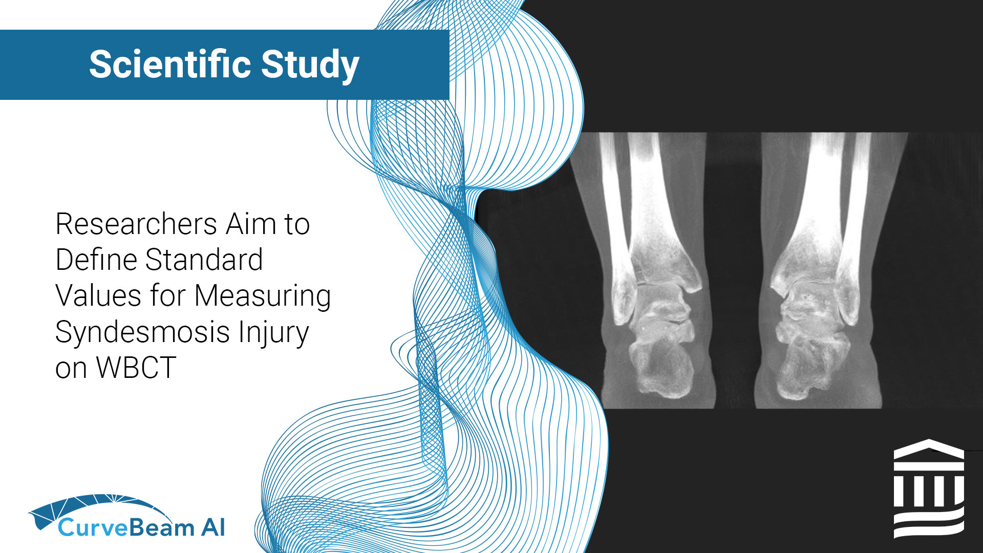
Researchers Aim to Define Standard Values for Measuring Syndesmosis Injury on WBCT
Key Points:
- Weight Bearing CT (WBCT) allows for bilateral comparative data that is imperative when diagnosing ankle syndesmosis.
- Side-to-side volume differences of 19% or greater may be indicative of an abnormality.
- Gender norms are important to consider when evaluating ankle syndesmosis.
The ankle syndesmosis is stabilized by a complex of ligaments, including the anterior inferior tibiofibular ligament, posterior inferior tibiofibular ligament, and the interosseous ligament and membrane. Syndesmotic injury is found in 25% of ankle sprain injuries and in 44% of ankle fractures. It’s necessary to distinguish the difference between injury and instability of syndesmosis because instability requires surgical intervention and is related to an increased risk of chronic pain and ankle joint arthritis.
While 3D measurement techniques using WBCT have gained popularity, normative bilateral comparative data still needs to be established. Dr Bejarano-Pineda et al out of the Foot & Ankle Research and Innovation Lab (FARIL) in the Department of Orthopaedic Surgery, Massachusetts General Hospital, Harvard Medical School in Boston, MA aimed to identify the side-to-side variations and gender differences in syndesmotic area and volume among individuals without syndesmotic injury, using WBCT.
Methods
Researchers conducted a retrospective analysis on bilateral ankle WBCT imaging of 88 individuals who underwent imaging for non-ankle related injuries or pathology. Two-dimensional area (at 1, 3, and 5cm proximal to the tibial plafond) and three-dimensional volumetric (from 0.5mm proximal to the tibial plafond and up to 3 and 5cm proximally) measurements were obtained for bilateral ankles. Mean values, percentage right-to-left differences and gender differences were all analyzed.
Results
It was found that there were significant differences between males and females in the area and volumetric measurements, with the largest differences observed for the volume from 0.5 to 5cm proximal to the tibial plafond. Statistically significant differences between genders were found for all area and volume measurements. In contrast, there were no significant differences found between laterality in 2D and 3D measurements among the entire cohort. The syndesmotic area at 5cm proximal to the tibial plafond showed the smallest difference between bilateral sides, with a 6.9% difference. The volume up to 5cm proximal to the tibial plafond showed the smallest change, with a 6.5% difference. None of the differences in laterality were statistically significant.
Conclusion
Researchers concluded that the mean side-to-side variation in the syndesmotic area and volume among individuals without syndesmotic injury was less than 9%, and a side-to-side volume difference greater than 19% might be indicative of abnormality. It was noted that gender-specific differences highlight the importance of considering gender norms in ankle syndesmosis evaluation and the need to use the contralateral side as a comparison.
To read the full study click here.
Related Posts


