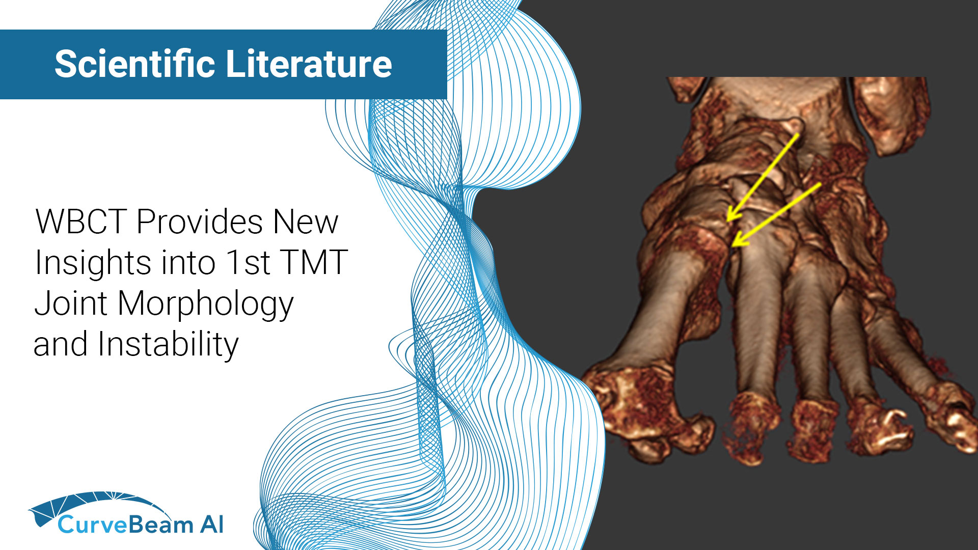
Key Points: First tarsometatarsal joint (TMT1) hypermobility is associated with hallux valgus (HV) which is…
FOOTInnovate Webinar Recap: Weight-Bearing CT as a Diagnosis Tool in Podiatric Radiology, Dr. Albert Armstrong DPM
Albert Armstrong, DPM, MS, BSRS, Professor of Radiology and Medical Director of Advanced Imaging at the Barry University School of Podiatric Medicine, is an important voice in the field of podiatry. He recently delivered a FOOTinnovate webinar titled, “Podiatric Radiology: Weight Bearing CT Imaging as an Essential Tool for Diagnosis.”
The Barry University Foot & Ankle Institute is comprised of three hospital-based podiatry clinics in greater Miami. Barry University podiatry students spend a portion of their third year rotating through these clinics. Barry University acquired a pedCAT system for both clinical and research applications in 2018.
In the webinar, Dr. Armstrong focused on how the CurveBeam pedCAT system improves diagnosis and workflow in clinic.
Click here to watch the FOOTInnovate webinar. A FOOTInnovate account is required.
pedCAT’s Efficiency
Dr. Armstrong explained a pedCAT study takes about the same amount of time and produces about the same amount of radiation as three traditional radiographs of the foot.
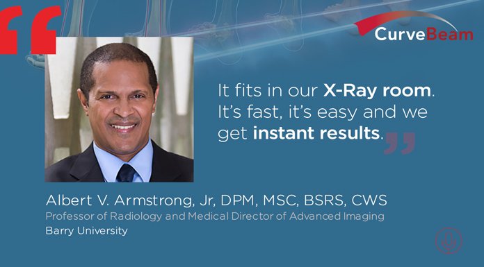
“The 3D, weight-bearing CT machine that we have is the pedCAT. … It’s small, compact, and it fits in our X-ray room,” Dr. Armstrong said. “It’s fast and easy, and we get instant results.” And patients often comment on the state-of-the-art technology.
pedCAT datasets can be displayed as Multiplanar Reformats (MPR), providing coronal, sagittal and transverse slices of the anatomy.
pedCAT users can also manually get “slices in virtually any plane,” Dr. Armstrong said.
Dr. Armstrong reviewed several cases from clinic in the webinar. Three of those cases are summarized below.
Case 1
Dr. Armstrong presented a case of a 52-year-old female who had undergone a bunionectomy three decades prior. She presented in clinic with pain in the left foot great toe. The pain was preventing the patient from walking, running and wearing heels.
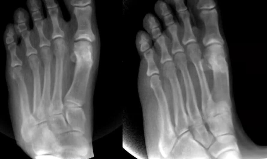
While traditional imagine didn’t provide a clear diagnosis, a coronal view created from the 3D volume revealed a large osteophyte and other factors that were hidden by the superimposition of the sesamoids and metatarsals in the X-Ray exam.
The more complete diagnosis, Dr. Armstrong said, led to a much better surgery plan for the patient.
The pedCAT and 3D volume imaging allows for detailed looks on several layers, as doctors can essentially “peel off,” as Dr. Armstrong puts it, layers ranging from shoes worn during weight-bearing scans to skin and soft tissue, etc.
Case 2
Another case, Dr. Armstrong said, highlights how critical weight-bearing imaging can be.
A 37-year-old male came to the Barry University Foot & Ankle Institute after a work injury was still painful two months later. The patient had fallen at his construction job. He went to the emergency room, where an X-Ray exam was read as negative, and he was diagnosed with an ankle sprain. The treatment prescribed was an ace wrap and a brace.
Two months later, the patient came to the Barry University Foot & Ankle Institute.
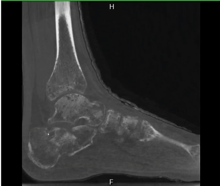
The fracture had been completely missed on the initial X-Ray. Patchy osteoporosis can be a sign of complex regional pain syndrome, and is typically radiographically indistinguishable as well.
Case 3
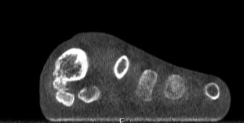
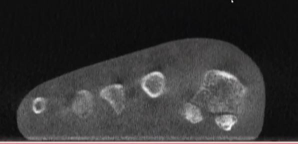
A 70-year-old male with a history of osteoarthritis and Type 2 Diabetes had synthetic cartilage implanted into his 1st MPJ joint 9 weeks prior. The patient had severe pain when standing and walking and had to walk on the lateral edge of his foot to compensate. Plain X-Rays came back negative.
With weight-bearing imaging, the coronal MPR images highlighted first metatarsal head was everted to such a degree that the patient was bearing weight on the tibial sesamoid when standing. That was causing his pain.
In summary, Dr. Armstrong said that pedCAT provides fast and efficient weight-bearing CT imaging, highlights the value of 3D volume and MPR images, and helps translate critical weight-bearing images into improved results for patients.
Related Posts