Weight bearing CT (WBCT) allows the patient to stand upright while a gantry rotates around…
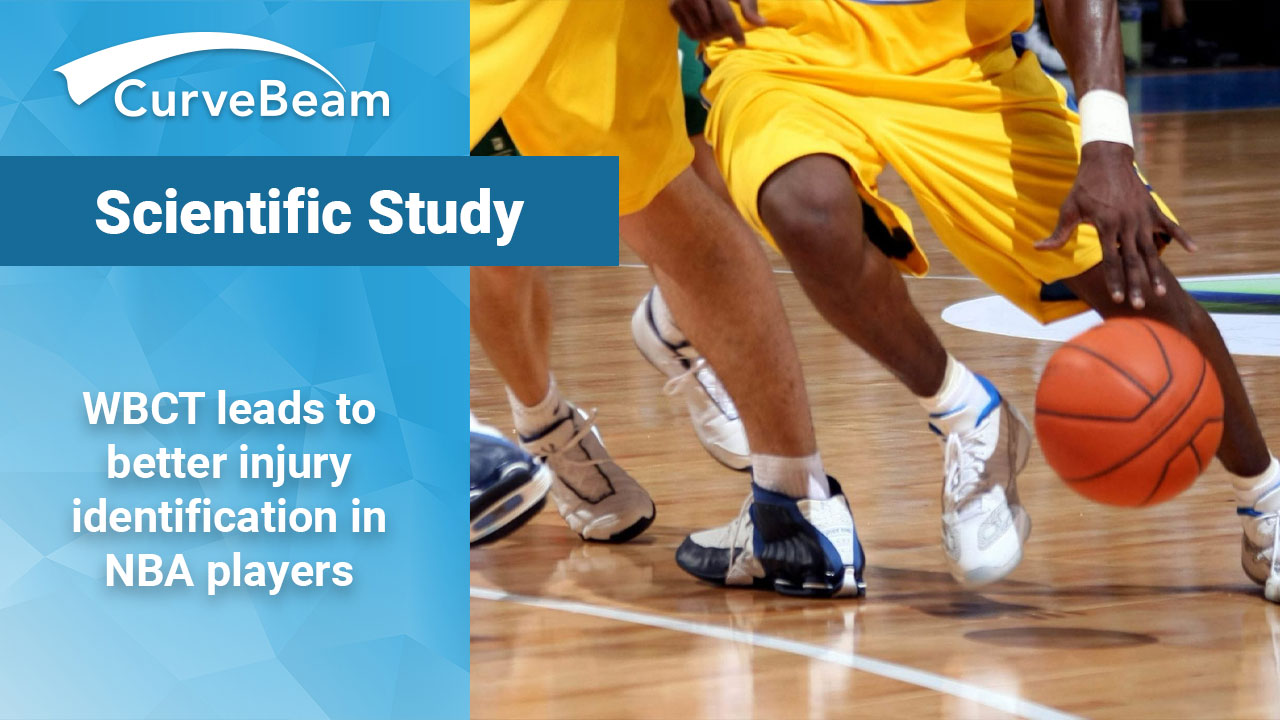
Study: Weight Bearing CT leads to better injury identification in NBA players
For professional athletes, injuries mean reduced playing time, impacted performance, and, in rare cases, an end to their careers. These injuries often affect the lower extremities, primarily because their sports require high-risk activities like jumping, cutting movements, and collision with other players. While traditional scanning techniques have mainly been used to identify injuries, a recent study found that weight-bearing cone beam computed tomography (CBCT) may be crucial to not only identify anatomic risks but also to help develop treatments explicitly tailored to the needs of professional athletes.
Incorporating new tech
In The Orthopaedic Journal of Sports Medicine, Dr. Cesar de Cesar Netto, et al. examined the morphology of foot injuries in 45 active NBA players. The doctors used weight-bearing CBCT scans to obtain 3D imaging of each foot. These scans provide more accurate alignment measurements than traditional scans and offer views of the foot while the player’s natural weight is being placed on it.
The study sought to discern whether the morphology of NBA players differed from that of the population at large, and whether the morphology changed based on position played. Foot and ankle injuries account for 27 percent of professional sports injuries, and 85 percent of basketball players experience at least one ankle sprain in their career.
Getting a better look
The players who participated in the study ranged from 20 to 31 years of age, and in total 29 right feet and 25 left feet were studied. All images were taken using a state-of-the-art pedCAT CurveBeam pedCAT system to obtain reliable and accurate images of each subject, and measurements were taken both manually and using the automatic TALAS measurement tool included with CurveBeam’s CubeVue software. TALAS is a research tool and is not available for clinical use. This is significant as it is the first time that a study of the foot morphotypes of NBA players has been conducted.
The study found that, for the most part, NBA players have standard alignment in their lower extremities, although they do tend to have high arches and varus hindfoot alignment. These trends were slight, but they are related to foot injuries and should still be noted. Building up a database of weight-bearing CBCT scans of professional athletes could also allow specialists to have a new control group to compare scans to, which would be enormously beneficial., the study authors said.
Better analysis means better results
Incorporating weight-bearing CBCT scans like those of CurveBeam’s pedCAT can save players, and the league as a whole, both time and money in the long term. Not only will they be able to watch for warning signs, but they will have a complete view of available injuries and will could develop more specific training regimens geared towards returning athletes to the court as quickly as possible.
You can read the full study by Dr. de Cesar Netto, et al. here.
Related Posts